. 2014 Nov 26:12:314.
doi: 10.1186/s12967-014-0314-y.

Reversal of severe angioproliferative pulmonary arterial hypertension and right ventricular hypertrophy by combined phosphodiesterase-5 and endothelin receptor inhibition

Affiliations

Reversal of severe angioproliferative pulmonary arterial hypertension and right ventricular hypertrophy by combined phosphodiesterase-5 and endothelin receptor inhibition

Maria A Cavasin et al. J Transl Med. .

Abstract

Background: Patients with pulmonary arterial hypertension (PAH) are treated with vasodilators, including endothelin receptor antagonists (ERAs), phosphodiesterase-5 (PDE-5) inhibitors, soluble guanylyl cyclase activators, and prostacyclin. Despite recent advances in pharmacotherapy for individuals with PAH, morbidity and mortality rates in this patient population remain unacceptably high. Here, we tested the hypothesis that combination therapy with two PAH drugs that target distinct biochemical pathways will provide superior efficacy relative to monotherapy in the rat SU5416 plus hypoxia (SU-Hx) model of severe angioproliferative PAH, which closely mimics the human condition.

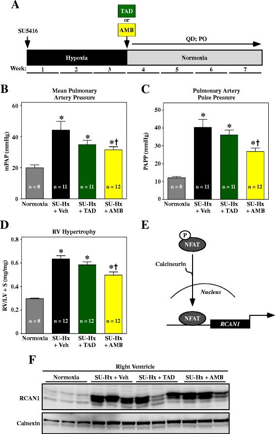
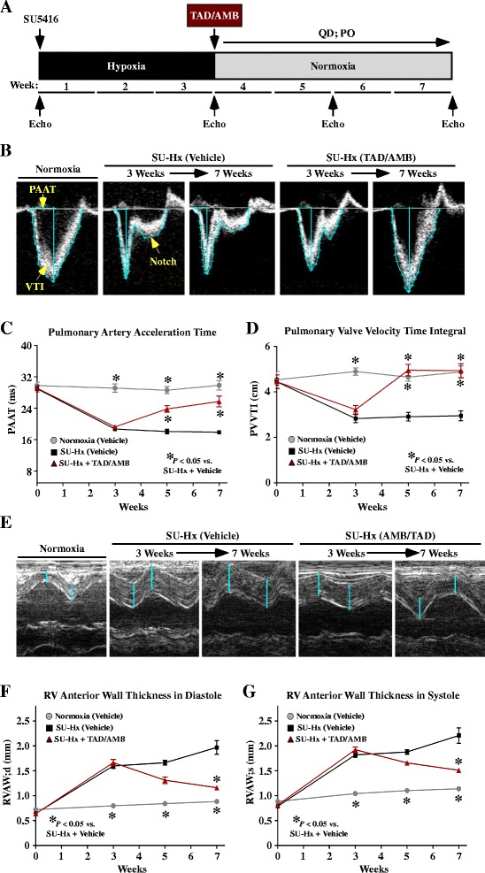
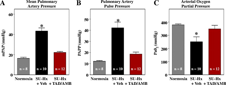
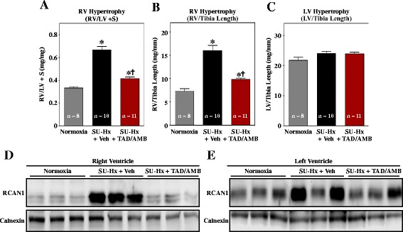
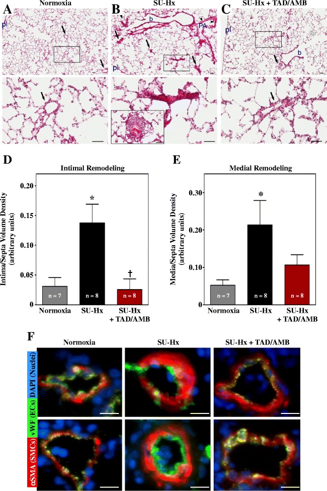
Methods: Male Sprague Dawley rats were injected with a single dose of SU5416, which is a VEGF receptor antagonist, and exposed to hypobaric hypoxia for three weeks. Rats were subsequently housed at Denver altitude and treated daily with the PDE-5 inhibitor, tadalafil (TAD), the type A endothelin receptor (ETA) antagonist, ambrisentan (AMB), or a combination of TAD and AMB for four additional weeks.

Results: Monotherapy with TAD or AMB led to modest reductions in pulmonary arterial pressure (PAP) and right ventricular (RV) hypertrophy. In contrast, echocardiography and invasive hemodynamic measurements revealed that combined TAD/AMB nearly completely reversed pulmonary hemodynamic impairment, RV hypertrophy, and RV functional deficit in SU-Hx rats. Efficacy of TAD/AMB was associated with dramatic reductions in pulmonary vascular remodeling, including suppression of endothelial cell plexiform lesions, which are common in human PAH.

Conclusions: Combined therapy with two vasodilators that are approved for the treatment of human PAH provides unprecedented efficacy in the rat SU-Hx preclinical model of severe, angioproliferative PAH.


Figures

Figure 1
Figure 1
Monotherapy to block PDE-5 or ET A signaling modestly reduces pulmonary pressure and RV hypertrophy. (A) Study design. Animals received 10 mg/kg tadalafil (TAD) or ambrisentan (AMB) once daily by oral gavage starting after week three. (B–C) Mean pulmonary arterial pressure (mPAP) and PA pulse pressure (PAPP) were measured invasively at study endpoint. (D) RV hypertrophy was assessed by weighing ventricular chambers at the time of necropsy, and is expressed as a ratio to LV + septum (S). (E) The gene encoding regulator of calcineurin-1 (RCAN1) is regulated by the nuclear factor of activated T cells (NFAT) transcription factor, which translocates to the nucleus in response to dephosphorylation by the pro-hypertrophic phosphatase, calcineurin. (F) RCAN1 expression in RV homogenates was detected by immunoblotting. Calnexin served as a loading control. Values represent mean +/−SEM. *P < 0.05 vs. normoxia; P < 0.05 vs. SU-Hx + vehicle (Veh). AMB significantly reduced pulmonary arterial pressure and RV hypertrophy, while TAD was without effect. Neither compound consistently blocked RV calcineurin signaling.
Figure 2
Figure 2
Combined PDE-5 and ET A inhibition reverses pulmonary hemodynamic impairment and RV hypertrophy in SU-Hx rats. (A) Study design. Animals received 10 mg/kg each of tadalafil (TAD) and ambrisentan (AMB) once daily by oral gavage starting after week three. (B) Pulmonary artery acceleration time (PAAT) and velocity time integral (VTI) were quantified using Doppler images. Systolic notching of PA blood flow in an SU-Hx rat treated with vehicle is indicated. (C and D) PAAT and VTI were significantly reduced in SU-Hx rats compared to normoxic controls, indicating increased pulmonary arterial pressure. PAAT and VTI were rescued by TAD/AMB treatment. (E–G) M-mode echocardiographic images revealed increased RV anterior wall thickness in SU-Hx rats, which was dramatically reduced by TAD/AMB treatment. For all graphs, values represent mean +/−SEM. *P < 0.05 vs. SU-Hx + vehicle (Veh). Normoxia (n = 8); SU-Hx (n = 10); SU-Hx + AMB/TAD (n = 12).
Figure 3
Figure 3
Combined PDE-5 and ET A inhibition suppresses pulmonary hypertension in SU-Hx rats. (A–B) Mean pulmonary arterial pressure (mPAP) and PA pulse pressure (PAPP) were measured invasively at study endpoint. (C) The partial pressure of oxygen in arterial blood was significantly increased by TAD/AMB. Values in all groups are higher than normal due to ventilation of animals with 100% O2 during pressure-volume analyses.
Figure 4
Figure 4
Combined PDE-5 and ET A inhibition improves RV function in SU-Hx rats. (A) Invasive pressure-volume analyses were performed at study endpoint. Representative RV pressure-volume loops are shown. The increase in RV end-systolic (B) and end-diastolic pressure (C) in SU-Hx rats was completely blocked by combined TAD/AMB treatment. (D) Pulmonary vascular resistance, calculated based on cardiac output, was lowered by TAD/AMB. (E) RV systolic function, based on ejection fraction (EF), was normalized by TAD/AMB. (F) RV cardiac output trended lower in SU-Hx rats, although the decrease was not statistically significant. *P < 0.05 vs. normoxia.
Figure 5
Figure 5
Combined PDE-5 and ET A inhibition blocks RV hypertrophy and pro-hypertrophic calcineurin signaling. RV hypertrophy, as determined by comparing weights of RV vs. LV + septum (S) (A) or RV mass to tibia length ratio (B) was significantly reduced by combined TAD/AMB treatment. (C) LV hypertrophy was not observed in the model. *P < 0.05 vs. normoxia; †P < 0.05 vs. SU-Hx + vehicle (D) Immunoblotting revealed normalization of RCAN1 expression in RVs of animals treated with TAD/AMB, indicating suppression of calcineurin signaling. Calnexin served as a loading control. (E) Elevated RCAN1 expression was observed in LVs of some SU-Hx rats.
Figure 6
Figure 6
Combined PDE-5 and ET A inhibition blocks pathological pulmonary vascular remodeling in SU-Hx rats. Images of a normoxic control lung (A), severely hypertensive lung in SU-Hx rats (B), and a lung from an SU-Hx animal treated with TAD/AMB for four weeks (C). Note that the SU-Hx lungs have marked muscularization of a pulmonary artery (PA, arrowheads), alongside a bronchiole (b), with several occlusive lesions located within the alveolar unit (arrows). TAD/AMB treatment led to normalization of pulmonary arteries, both at the bronchiolar level (b) as well as in intra-alveolar compartment (arrows), similar to the pulmonary artery morphology (arrows) seen in control lungs. High magnification images of the boxed areas are shown in the lower panels. In addition, in (B) the lower panel shows a classical plexiform-like lesion in a vehicle treated SU-Hx rat. This image was taken from a different slide. pl, visceral pleura; magnification bar =50 μm. Intimal occlusion (D) and medial thickening (E) in SU-Hx rats was reduced by TAD/AMB treatment. Values represent mean +/−SEM. *P < 0.05 vs. normoxia. †P < 0.05 vs. SU-Hx + vehicle. (F) Representative images of lung arterioles stained for von Willebrand factor (vWF; endothelial cell marker) and alpha smooth muscle actin (αSMA; smooth muscle cell marker). Images are overlaid with DAPI-stained nuclei; magnification bar =10 μm. Vessels from SU-Hx rats exhibited expansion of smooth muscle and endothelial cells, which was reduced by TAD/AMB treatment.

References

    1. Humbert M, Morrell NW, Archer SL, Stenmark KR, MacLean MR, Lang IM, Christman BW, Weir EK, Eickelberg O, Voelkel NF, Rabinovitch M. Cellular and molecular pathobiology of pulmonary arterial hypertension. J Am Coll Cardiol. 2004;43:13S–24S. doi: 10.1016/j.jacc.2004.02.029. - DOI - PubMed
    1. Hoeper MM, Barst RJ, Bourge RC, Feldman J, Frost AE, Galie N, Gomez-Sanchez MA, Grimminger F, Grunig E, Hassoun PM, Morrell NW, Peacock AJ, Satoh T, Simonneau G, Tapson VF, Torres F, Lawrence D, Quinn DA, Ghofrani HA. Imatinib mesylate as add-on therapy for pulmonary arterial hypertension: results of the randomized IMPRES study. Circulation. 2013;127:1128–1138. doi: 10.1161/CIRCULATIONAHA.112.000765. - DOI - PubMed
    1. Wu Y, O’Callaghan DS, Humbert M. An update on medical therapy for pulmonary arterial hypertension. Curr Hypertens Rep. 2013;15:614–622. doi: 10.1007/s11906-013-0394-8. - DOI - PubMed
    1. Stenmark KR, Fagan KA, Frid MG. Hypoxia-induced pulmonary vascular remodeling: cellular and molecular mechanisms. Circ Res. 2006;99:675–691. doi: 10.1161/01.RES.0000243584.45145.3f. - DOI - PubMed
    1. Stenmark KR, Meyrick B, Galie N, Mooi WJ, McMurtry IF. Animal models of pulmonary arterial hypertension: the hope for etiological discovery and pharmacological cure. Am J Physiol Lung Cell Mol Physiol. 2009;297:L1013–L1032. doi: 10.1152/ajplung.00217.2009. - DOI - PubMed

Publication types

Substances