Review
. 2015 Feb;35(2):176-85.
doi: 10.1038/jcbfm.2014.206. Epub 2014 Nov 26.

Lactate transport and signaling in the brain: potential therapeutic targets and roles in body-brain interaction

Affiliations
Review

Lactate transport and signaling in the brain: potential therapeutic targets and roles in body-brain interaction

Linda Hildegard Bergersen. J Cereb Blood Flow Metab. 2015 Feb.

Abstract

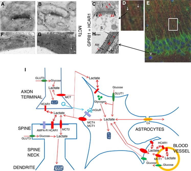
Lactate acts as a 'buffer' between glycolysis and oxidative metabolism. In addition to being exchanged as a fuel by the monocarboxylate transporters (MCTs) between cells and tissues with different glycolytic and oxidative rates, lactate may be a 'volume transmitter' of brain signals. According to some, lactate is a preferred fuel for brain metabolism. Immediately after brain activation, the rate of glycolysis exceeds oxidation, leading to net production of lactate. At physical rest, there is a net efflux of lactate from the brain into the blood stream. But when blood lactate levels rise, such as in physical exercise, there is net influx of lactate from blood to brain, where the lactate is used for energy production and myelin formation. Lactate binds to the lactate receptor GPR81 aka hydroxycarboxylic acid receptor (HCAR1) on brain cells and cerebral blood vessels, and regulates the levels of cAMP. The localization and function of HCAR1 and the three MCTs (MCT1, MCT2, and MCT4) expressed in brain constitute the focus of this review. They are possible targets for new therapeutic drugs and interventions. The author proposes that lactate actions in the brain through MCTs and the lactate receptor underlie part of the favorable effects on the brain resulting from physical exercise.


Figures

Figure 1
Figure 1
Lactate transport and signaling in the brain. (A) Electron micrograph showing double immunogold labeling at synapses between parallel fiber terminals (T) and Purkinje cell spines (S). The section was double-labeled with antibodies to GluR2/3 (15 nm gold particles) and to MCT2 (10 nm gold particles). Note colocalization of MCT2 with GluR2/3 at the synaptic membrane and intracellularly in the spine. Cerebellar cortex, molecular layer. (B) The membranes of the Bergmann glia (A) facing the parallel fiber terminals (T) and Purkinje cell spines (S) are labeled with antibody to MCT4 (10 nm gold particles). (C) The lactate receptor (10 nm gold, red arrowheads) is at the postsynaptic membrane (black arrowheads). Synapse of a nerve terminal (t) on a dendritic spine (s). Stratum radiatum, hippocampus CA1. (D, E) The lactate receptor GPR81/HCAR1 (green) is in the pyramidal cell somatodendritic compartment including spines (white arrows in D), and in microvascular endothelium (white v in E). (D) Magnification of area framed in (E) is shown. Hippocampus CA1. Neurons labeled for microtubule-associated protein 2 (MAP2) (red), nuclei with DAPI (blue). (F) The endfeet (Ae) of the Bergmann glia facing the endothelium (E) strongly express MCT4 (10 nm gold particles). (G) MCT1 (10 nm gold particles) is in the endothelium facing the lumen of the capillary, including in itracellular vesicular organelles (partly indicated by white arrows). (H) Electron micrograph showing GPR81 immunogold particles (red arrowheads) at luminal (short arrow) and abluminal (Abl) membrane of the vascular endothelium and at the perivascular astrocytic end-foot membrane (Ae) at a hippocampal blood vessel. (I) Schematic representation of L-lactate production and action at the ‘tripartite synapse' of axon, dendrite, and astrocyte, and at the glio-vascular junction (see text). Lactate, formed by glycolysis in brain cells or entering from blood, migrates down concentration gradients (of lactate and cotransported proton) between intracellular and extracellular compartments of neurons, astrocytes, and endothelial cells (orange circle), catalyzed by monocarboxylate transporters (MCT1, MCT2, and MCT4, red ovals). Lactate also migrates along the extracellular space, as well as throughout the astrocytic syncytial network via connexin gap junctions (Cx, orange oval). For simplification, pyruvate and other intermediates are not shown. HCAR1 (red rectangles), lactate receptors, the lactate binding site facing the extracellular space. GLUT1 and GLUT3 (green ovals), glucose transporters, providing glucose for brain cells. Positions of ionotropic glutamate receptors (AMPA-R, purple rectangles) and metabotropic glutamate receptors (blue serpents), influencing and influenced by lactate, are indicated. Mitochondria (dark-blue symbols) shun dendritic spines and thin astroglial processes. Astroglial processes contain glycogen particles (green spheres). (A, B, F, G modified from Bergersen and C, D, E, H modified from/based on Lauritzen.)

References

    1. Bergersen LH. Is lactate food for neurons? Comparison of monocarboxylate transporter subtypes in brain and muscle. Neuroscience. 2007;145:11–19. - PubMed
    1. Halestrap AP. The SLC16 gene family—structure, role and regulation in health and disease. Mol Aspects Med. 2013;34:337–349. - PubMed
    1. Bergersen LH, Gjedde A. Is lactate a volume transmitter of metabolic states of the brain. Front Neuroenergetics. 2012;4:5. - PMC - PubMed
    1. Lauritzen KH, Morland C, Puchades M, Holm-Hansen S, Hagelin EM, Lauritzen F, et al. Lactate receptor sites link neurotransmission, neurovascular coupling, and brain energy metabolism. Cereb Cortex. 2013;24:2784–2795. - PubMed
    1. Brooks GA. Cell-cell and intracellular lactate shuttles. J Physiol. 2009;587:5591–5600. - PMC - PubMed

Publication types

MeSH terms

Substances