. 2015 Jan 16;290(3):1559-69.
doi: 10.1074/jbc.M114.592246. Epub 2014 Nov 25.

Use of chimeras, point mutants, and molecular modeling to map the antagonist-binding site of 4,4',4″,4‴-(carbonylbis-(imino-5,1,3-benzenetriylbis(carbonylimino)))tetrakisbenzene-1,3-disulfonic acid (NF449) at P2X1 receptors for ATP

Affiliations

Use of chimeras, point mutants, and molecular modeling to map the antagonist-binding site of 4,4',4″,4‴-(carbonylbis-(imino-5,1,3-benzenetriylbis(carbonylimino)))tetrakisbenzene-1,3-disulfonic acid (NF449) at P2X1 receptors for ATP

Louise K Farmer et al. J Biol Chem. .

Abstract

P2X receptor subtype-selective antagonists are promising candidates for treatment of a range of pathophysiological conditions. However, in contrast to high resolution structural understanding of agonist action in the receptors, comparatively little is known about the molecular basis of antagonist binding. We have generated chimeras and point mutations in the extracellular ligand-binding loop of the human P2X1 receptor, which is inhibited by NF449, suramin, and pyridoxal-phosphate-6-azophenyl-2,4-disulfonate, with residues from the rat P2X4 receptor, which is insensitive to these antagonists. There was little or no effect on sensitivity to suramin and pyridoxal-phosphate-6-azophenyl-2,4-disulfonate in chimeric P2X1/4 receptors, indicating that a significant number of residues required for binding of these antagonists are present in the P2X4 receptor. Sensitivity to the P2X1 receptor-selective antagonist NF449 was reduced by ∼60- and ∼135-fold in chimeras replacing the cysteine-rich head, and the dorsal fin region below it in the adjacent subunit, respectively. Point mutants identified the importance of four positively charged residues at the base of the cysteine-rich head and two variant residues in the dorsal fin for high affinity NF449 binding. These six residues were used as the starting area for molecular docking. The four best potential NF449-binding poses were then discriminated by correspondence with the mutagenesis data and an additional mutant to validate the binding of one lobe of NF449 within the core conserved ATP-binding pocket and the other lobes coordinated by positive charge on the cysteine-rich head region and residues in the adjacent dorsal fin.

Keywords: ATP; Electrophysiology; Ion Channel; Ligand-gated Ion Channel; Mutagenesis; NF449; P2X Receptor; PPADS; Purinergic Receptor; Suramin.


Figures

FIGURE 1.
FIGURE 1.
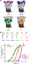
Characterization of ATP at chimeric P2X receptors. a, location of regions A–D on an ATP-bound homology model of the P2X1 receptor. Regions A–D are shown in color; ATP is shown in yellow. Transmembrane domains are shown in dark gray. b, upper, representative traces for WT and chimeric receptors at maximal ATP concentrations. The bars represent a 3-s ATP application. Traces have been normalized to peak currents to allow for comparison. Lower, concentration-response curves for ATP. Black asterisks indicate significant shifts in EC50 from the WT P2X1 receptor, red asterisks from the P2X4 receptor, and blue asterisks from both receptors. ****, p < 0.0001.
FIGURE 2.
FIGURE 2.
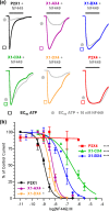
NF449 action at chimeric P2X receptors. a, representative traces showing the effect of NF449 (10 nm) on currents evoked by an EC90 of ATP in Xenopus oocytes expressing WT and chimeric receptors. The bars indicate a 3-s agonist/antagonist application. b, NF449 inhibition curves at an EC90 of ATP. Asterisks indicate a significant shift in IC50 from the hP2X1 receptor. *, p < 0.05; ****, p < 0.0001.
FIGURE 3.
FIGURE 3.
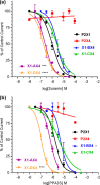
Suramin and PPADS antagonism at chimeric P2X receptors. a, comparison of suramin action in WT receptors and chimeras. b, PPADS inhibition curves for chimeric and WT receptors. In all cases, antagonist action was determined against an EC90 of ATP. Asterisks indicate a significant shift from the WT P2X1 receptor. **, p < 0.05; ***, p < 0.0001.
FIGURE 4.
FIGURE 4.
Introduction of four charges reintroduced NF449 sensitivity to the X1-BX4 chimera. a, location of Lys-136, Lys-138, Arg-139, and Lys-140 in a P2X1 receptor homology model. Docked ATP is shown in yellow. b, NF449 inhibition curves showing the effect of reintroducing the positive charges to the X1-BX4 chimera and NF449 action in the P2X4(4+) receptor. Asterisks indicate a significant difference in IC50 compared with the P2X1 receptor. ****, p < 0.0001.
FIGURE 5.
FIGURE 5.
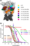
Effects of NF449 at region C subchimeras. a, location of residues swapped to generate chimeras shown in a homology model of the P2X1 receptor. Docked ATP is shown in yellow, and transmembrane domains are shown in dark gray. b, NF449 inhibition curves for subchimeras. Curves were generated at an EC90 of ATP. Asterisks represent a significant difference in IC50 compared with the P2X1 receptor. ****, p < 0.0001.
FIGURE 6.
FIGURE 6.
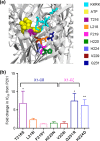
Effects of NF449 at point mutants. a, location of point mutated residues in a P2X1 receptor homology model. Docked ATP is shown in yellow. Residues were mutated to the equivalent residue of the P2X4 receptor. b, histogram showing the -fold change in NF449 inhibition compared with the WT P2X1 receptor at an EC90 of ATP. Asterisks represent a significant difference. *, p < 0.05; **, p < 0.01.
FIGURE 7.
FIGURE 7.
Docking poses for the NF449-P2X1 complex. a and b, overlay of the four docking poses A–D on the P2X1 receptor shown from different angles. The P2X1 receptor is shown in surface representation, highlighting the positively charged residues of the ATP-binding site (Lys-68, Lys-70, Arg-292, and Lys-309) in light blue, the positively charged residues of the cysteine-rich head region (Lys-136, Lys-138, Arg-139, and Lys-140) in dark blue, Thr-216 and Gln-231 in green, region X1-CγX4 (residues 210–215; see “Results”) in pale cyan, and His-224 in purple. NF449 poses A (red), B (orange), C (pink), and D (yellow) are shown as a mixture of sphere representation (for the core of the poses) and stick representation (for the arms of the poses). All four poses bind to the cleft between the cysteine-rich head region, ATP-binding site, and dorsal fin. c, molecular structure of the NF449 sodium salt. d, snapshot of NF449 from the pose C trajectory. NF449 and residues Lys-68, Lys-70, Lys-136, Lys-138, Arg-139, Lys-140, Thr-216, His-224, Gln-231, Arg-292, and Lys-309 are shown in stick representation. e, distances between NF449 sulfonate sulfur atoms and His-224 Nϵ (black), Thr-216 Oγ (green), and the amide nitrogen of Gln-231 monitored over the 10-ns molecular dynamics simulation of pose C. NF449/His-224 and NF449/Thr-216 interactions are present in most of the frames, and NF449/Gln-231 interactions are less stable in this simulation and are present only between 1.5 and 4.5 ns. f, section of NF449 pose C interacting with the ATP-binding site. Potential salt bridges are indicated by dashed black lines. g, section of NF449 pose A and residues Lys-136, Lys-138, and Arg-139 of the cysteine-rich head region. h, arm of NF449 pose D forming H-bonds with Thr-216 and Gln-231 side chains.

References

    1. Burnstock G. (2012) Purinergic signalling: its unpopular beginning, its acceptance and its exciting future. BioEssays 34, 218–225 - PubMed
    1. Valera S., Hussy N., Evans R. J., Adami N., North R. A., Surprenant A., Buell G. (1994) A new class of ligand-gated ion channel defined by P2X receptor for extracellular ATP. Nature 371, 516–519 - PubMed
    1. Brake A. J., Wagenbach M. J., Julius D. (1994) New structural motif for ligand-gated ion channels defined by an ionotropic ATP receptor. Nature 371, 519–523 - PubMed
    1. Kaczmarek-Hájek K., Lörinczi E., Hausmann R., Nicke A. (2012) Molecular and functional properties of P2X receptors–recent progress and persisting challenges. Purinergic Signal. 8, 375–417 - PMC - PubMed
    1. Surprenant A., North R. A. (2009) Signaling at purinergic P2X receptors. Annu. Rev. Physiol. 71, 333–359 - PubMed

Publication types

Substances

Associated data

LinkOut - more resources