. 2014 Nov 20;16(11):972-81.
doi: 10.1016/j.neo.2014.09.008. eCollection 2014 Nov.

E7080 (lenvatinib), a multi-targeted tyrosine kinase inhibitor, demonstrates antitumor activities against colorectal cancer xenografts

Affiliations

E7080 (lenvatinib), a multi-targeted tyrosine kinase inhibitor, demonstrates antitumor activities against colorectal cancer xenografts

Armin Wiegering et al. Neoplasia. .

Abstract

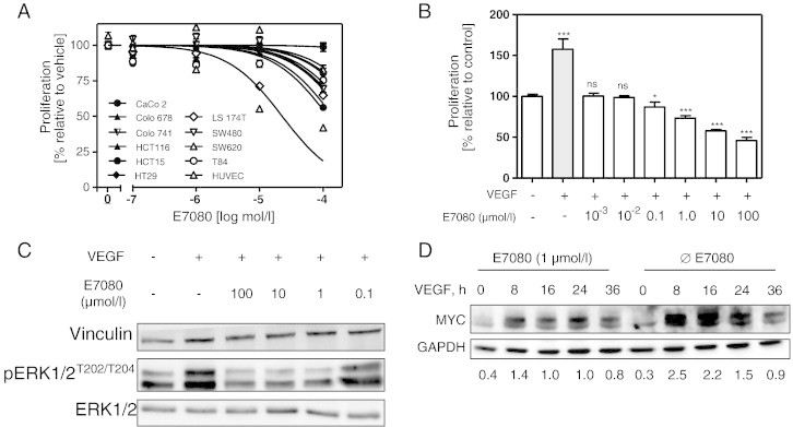
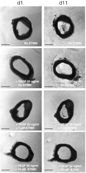
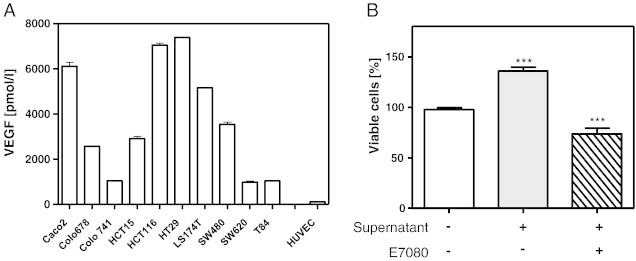
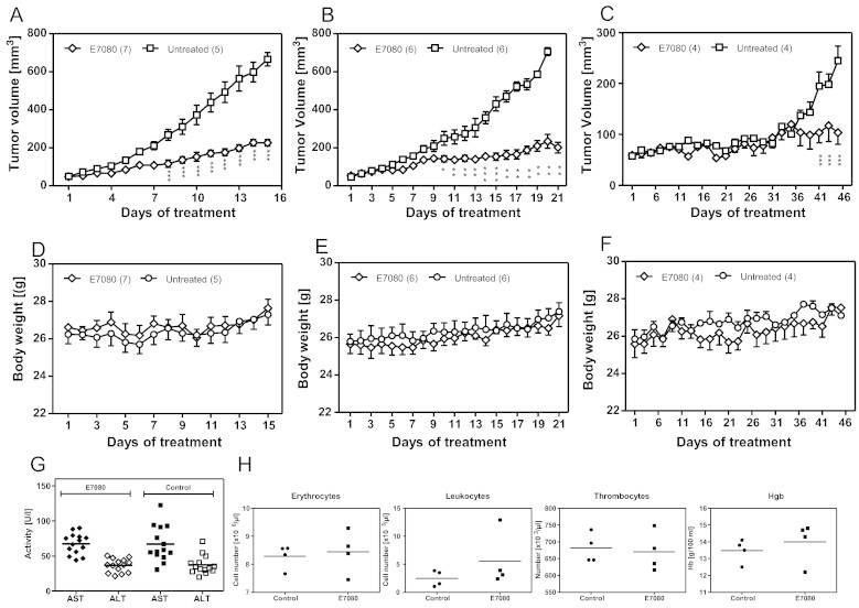
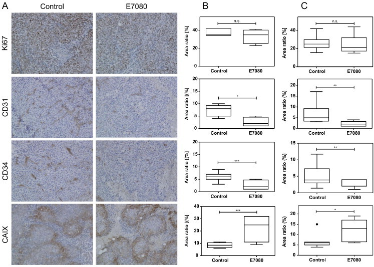
Clinical prognosis of metastasized colorectal carcinoma (CRC) is still not at desired levels and novel drugs are needed. Here, we focused on the multi-tyrosine kinase inhibitor E7080 (Lenvatinib) and assessed its therapeutic efficacy against human CRC cell lines in vitro and human CRC xenografts in vivo. The effect of E7080 on cell viability was examined on 10 human CRC cell lines and human endothelial cells (HUVEC). The inhibitory effect of E7080 on VEGF-induced angiogenesis was studied in an ex vivo mouse aortic ring angiogenesis assay. In addition, the efficacy of E7080 against xenografts derived from CRC cell lines and CRC patient resection specimens with mutated KRAS was investigated in vivo. A relatively low cytotoxic effect of E7080 on CRC cell viability was observed in vitro. Endothelial cells (HUVEC) were more susceptible to the incubation with E7080. This is in line with the observation that E7080 demonstrated an anti-angiogenic effect in a three-dimensional ex vivo mouse aortic ring angiogenesis assay. E7080 effectively disrupted CRC cell-mediated VEGF-stimulated growth of HUVEC in vitro. Daily in vivo treatment with E7080 (5 mg/kg) significantly delayed the growth of KRAS mutated CRC xenografts with decreased density of tumor-associated vessel formations and without tumor regression. This observation is in line with results that E7080 did not significantly reduce the number of Ki67-positive cells in CRC xenografts. The results suggest antiangiogenic activity of E7080 at a dosage that was well tolerated by nude mice. E7080 may provide therapeutic benefits in the treatment of CRC with mutated KRAS.


Figures

Figure 1
Figure 1
Effects of E7080 on cell viability and cell signalling. (A) Dose-response effects of E7080 on cell viability of human colorectal carcinoma (CRC) cells and endothelial cells (HUVEC). IC50 values were determined in serum reduced (1%) RPMI 1640 medium following treatment with different concentrations of E7080 for 72 hours. The final concentration of DMSO (vehicle) was ≤ 1%. Results are expressed as the mean ± S.E.M. of at least three independent proliferation assays with hexaplicates. IC50 values were determined using GraphPad Prism software. See also Table 1. (B) E7080 inhibits VEGF-induced HUVEC proliferation. HUVEC (1.5 × 10⁴ cells) were seeded in 96-well plates coated with attachment factor (#S-006-100, Gibco Life Technologies) for 24 hours. The culture medium was changed and HUVEC were incubated in serum reduced (1%) RPMI 1640 medium in the presence of VEGF (20 ng/ml) and different concentrations of E7080 as indicated. Cell viability was determined with WST-8 assay after 48 hours. Shown are the results of one representative experiment with hexaplicates. *P < 0.05, **P < 0.01, ***P < 0.001 to HUVEC control proliferation (without VEGF and E7080). (C) E7080 inhibits VEGF-induced phosphorylation of ERK1/2. Serum starved HUVEC were incubated with indicated amounts of E7080 for 4 hours and then stimulated with VEGF (20 ng/ml) for 5 minutes. Cells were harvested immediately on ice and immunoblotted with indicated antibodies against ERK1/2 and pERK1/2 as described in Materials and Methods. (D) VEGF-stimulated MYC expression is suppressed by E7080. HUVEC were incubated in serum reduced (1%) RPMI 1640 medium with E7080 (1 μmol/l) for 4 hours and afterwards with VEGF (20 ng/ml) until indicated time points; subsequently cells were harvested for western blot analysis of MYC. Detection of MYC and GAPDH was performed using enzyme-linked chemiluminescence. GAPDH was used as loading control. The intensity of the signals corresponding to MYC and GAPDH was quantified by densitometry using ImageJ software (National Institutes of Health). The level of MYC protein is expressed as the ratio of the MYC band to that of GAPDH of a representative experiment.
Figure 2
Figure 2
E7080 inhibits VEGF-induced micro vessel sprouting in three-dimensional ex vivo mouse aortic ring angiogenesis assay. Angiogenesis was induced with 50 ng/ml VEGF and E7080 was used at the indicated final concentrations. Photographs were taken at day 1 and day 11. Representative photographs are shown. Scale bar = 250 μm.
Figure 3
Figure 3
Colorectal carcinoma (CRC) cell-secreted VEGF induces human endothelial cell (HUVEC) proliferation. (A) Presence of VEGF in the supernatant of CRC cell cultures. VEGF was detected with specific ELISA according to manufacturer’s instructions (R&D Systems GmbH, Germany). For this, 1 million CRC cells and HUVEC were cultured in 6-well plates with serum reduced (1%) RPMI 1640 medium for 24 hours. Levels of detected VEGF ranged from 1000 pmol/l for T84 to over 7000 pmol/l for HT29. In supernatant of HUVEC, detected VEGF levels ranged below 100 pmol/l. (B) E7080 suppresses HUVEC proliferation in the presence of CRC cell-secreted VEGF. Representative results shown for CaCo2 cells with CRC cell supernatant and/or E7080 (1 μmol/l). See also Supplementary Figure 2. HUVEC (1.5 × 10⁴ cells) were seeded in 96-well plates coated with attachment factor (#S-006-100, Gibco Life Technologies) for 24 hours. The cells were washed with PBS and incubated with 100 μl CRC cell line supernatant, 100 μl fresh serum-reduced (1%) RPMI 1640 medium and E7080 (0.1 μmol/l) for 72 hours. Cell viability was determined with WST-8 assay. ***P < 0.001 to HUVEC control proliferation (without CRC cell supernatant and E7080). (B) E7080 suppresses HUVEC proliferation in the presence of CRC cell-secreted VEGF. Representative results shown for CaCo2 cells with CRC cell supernatant and/or E7080 (1 μmol/l). See also Supplementary Figure 2. HUVEC (1.5 × 10⁴ cells) were seeded in 96-well plates coated with attachment factor (#S-006-100, Gibco Life Technologies) for 24 hours. The cells were washed with PBS and incubated with 100 μl CRC cell line supernatant, 100 μl fresh serum-reduced (1%) RPMI 1640 medium and E7080 (0.1 μmol/l) for 72 hours. Cell viability was determined with WST-8 assay. ***P < 0.001 to HUVEC control proliferation (without CRC cell supernatant and E7080).
Figure 4
Figure 4
E7080 retards tumor growth in vivo without adverse events. Xenografts of LS174T (A), HCT116 (B) and patient resection specimens (C). Nude mice received daily 5 mg/kg E7080 orally or vehicle control. (D-F) Body weight was monitored throughout the indicated period. At the end of experiment the activity of liver enzymes alanine transaminase, AST, and aspartate transaminase, ALT (G) and blood parameters (H) were estimated. Values represent means with bars indicating standard errors. *P < 0.05, **P < 0.01, ***P < 0.001 to untreated control.
Figure 5
Figure 5
The E7080-mediated influence of microvascularization induces tumor hypoxia. (A) Representative immunohistochemical staining of untreated and E7080 treated tumors (LS174T). Quantitative analysis of LS174T derived xenografts (B) and HCT116 derived xenografts (C), both KRAS mutated. Tumor sections were stained towards Ki67, CD31, CD34, and CAIX (carbonic anhydrase 9) using standard immunohistochemical procedures as described in Material and Methods. Results are shown as Tukey boxplot with first, second (the median) and third quartiles of four to seven animals (Figure 4) per group. The lower whisker represents the 1.5 interquartile range (IQR) of the lower quartile, and the top whisker represents the 1.5 IQR of the upper quartile. The point indicates an outlier. *P < 0.05, **P < 0.01, ***P < 0.001 to control tumors. For the results of CRC xenografts derived from patient resection specimens with KRAS mutation see Supplementary Figure 3. (A) Representative immunohistochemical staining of untreated and E7080 treated tumors (LS174T). Quantitative analysis of LS174T derived xenografts (B) and HCT116 derived xenografts (C), both KRAS mutated. Tumor sections were stained towards Ki67, CD31, CD34, and CAIX (carbonic anhydrase 9) using standard immunohistochemical procedures as described in Material and Methods. Results are shown as Tukey boxplot with first, second (the median) and third quartiles of four to seven animals (Figure 4) per group. The lower whisker represents the 1.5 interquartile range (IQR) of the lower quartile, and the top whisker represents the 1.5 IQR of the upper quartile. The point indicates an outlier. *P < 0.05, **P < 0.01, ***P < 0.001 to control tumors. For the results of CRC xenografts derived from patient resection specimens with KRAS mutation see Supplementary Figure 3.

References

    1. Siegel R., Desantis C., Jemal A. Colorectal cancer statistics, 2014. CA Cancer J Clin. 2014;64(2):104–117. - PubMed
    1. Lorenz M., Staib-Sebler E., Hochmuth K., Heinrich S., Gog C., Vetter G., Encke A., Muller H.H. Surgical Resection of Liver Metastases of Colorectal Carcinoma: Short and Long-Term Results. Semin Oncol. 2000;27(5 Suppl. 10):112–119. - PubMed
    1. Folkman J. Angiogenesis in cancer, vascular, rheumatoid and other disease. Nat Med. 1995;1(1):27–31. - PubMed
    1. Goel H.L., Mercurio A.M. VEGF targets the tumour cell. Nat Rev Cancer. 2013;13(12):871–882. - PMC - PubMed
    1. Cunningham D., Humblet Y., Siena S., Khayat D., Bleiberg H., Santoro A., Bets D., Mueser M., Harstrick A., Verslype C. Cetuximab monotherapy and cetuximab plus irinotecan in irinotecan-refractory metastatic colorectal cancer. N Engl J Med. 2004;4:337–345. - PubMed

Publication types

MeSH terms