Review
. 2014 May 31;8(5):1-8.
doi: 10.3941/jrcr.v8i5.1721. eCollection 2014 May.

Delayed leukoencephalopathy after acute carbon monoxide intoxication

Affiliations
Review

Delayed leukoencephalopathy after acute carbon monoxide intoxication

Ana Filipa Geraldo et al. J Radiol Case Rep. .

Abstract

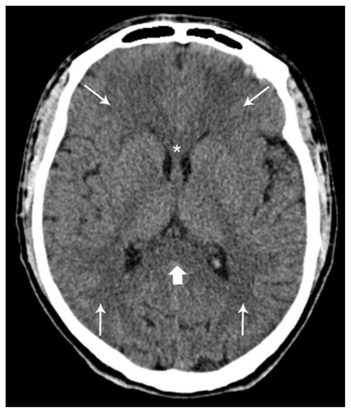
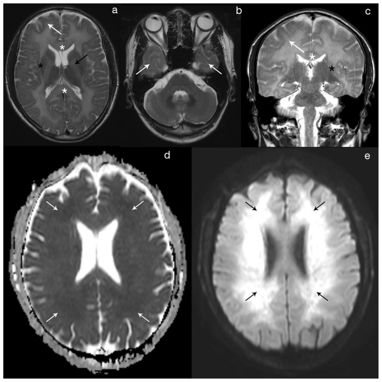
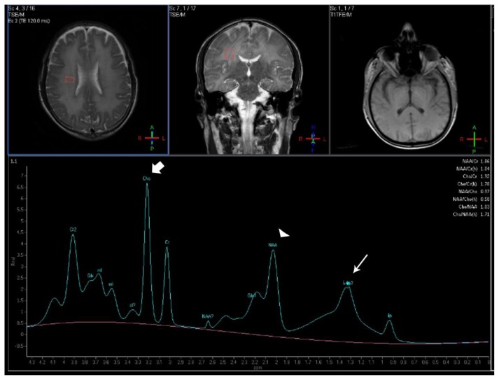
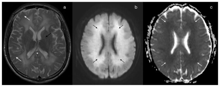
Delayed leukoencephalopathy is an uncommon complication of hypoxic-ischemic events of different etiologies, including carbon monoxide intoxication. We present a case of a 40-year-old male patient who was admitted with rapidly progressive neurocognitive and behavioral deficits. There was a history of accidental carbon monoxide intoxication one month before, presenting with loss of consciousness and short hospitalization, followed by a complete clinical recovery. The imaging studies in the delayed phase depicted confluent, symmetric supra-tentorial white matter lesions in keeping with diffuse demyelinization. Restricted diffusion and metabolite abnormalities in magnetic resonance proton spectroscopy were also seen. The diagnosis of CO-mediated delayed post-hypoxic leukoencephalopathy was assumed after exclusion of other mimickers. Hyperbaric oxygen therapy was tentatively performed and the patient had a favorable clinical and radiological evolution.

Keywords: Carbon monoxide poisoning; delayed post-ischemic encephalopathy; diffusion magnetic resonance imaging; heroin inhalation syndrome; magnetic resonance spectroscopy; neurotoxicity syndromes.


Figures

Figure 1
Figure 1
Forty-year-old male with diffuse post-hypoxic demyelination (1 month after the initial event and before treatment). FINDINGS: Axial non-contrast CT scan shows diffuse hypodensity in the periventricular white matter (small arrows), the splenium (large arrow) and the genu (asterisks) of the corpus callosum. TECHNIQUE: Phillips Brilliance 64; FOV 247; 2 mm slice thickness; 120 Kv; 238 mA; 399 mAs.
Figure 2
Figure 2
Forty-year-old male with diffuse post-hypoxic demyelination (1 month after the initial event and before treatment). FINDINGS: Axial T2 (2a) and coronal T2 (2c) show a diffuse, confluent hyperintensity involving predominantly the periventricular and deep white-matter, as well as the corpus callosum (white asterisks), the internal (black arrow) and the external (black asterisks) capsules. There is also some extension of the signal abnormality to the sub-cortical white-matter (white arrow). The cortex and the deep gray-matter are spared. Axial T2 (2b) at one inferior level depicts the involvement of the anterior temporal sub-cortical white-matter (arrows), with normal findings in the pons and cerebellum. Axial DWI (2e) shows diffuse hyperintensity in the peri-ventricular and deep white-matter with corresponding hypointensity on the axial ADC map (2d) reflecting restricted diffusion. TECHNIQUE: Philips Intera 3T; Axial T2 (TR 2000/TE 80; Flip angle 90; FOV 230; slice thickness 5mm; NEX 1); Coronal T2 (TR 3000/TE 80; Flip angle 90; FOV 220; slice tickness 4mm; NEX 1); Axial DWI and ADC (TR 3869.16/TE 58.714; Flip angle 18; FOV 240; b value 1000; NEX 2).
Figure 3
Figure 3
Forty-year-old male with diffuse post-hypoxic demyelination (one month after the initial event and pre-treatment). FINDINGS: The MRS spectrum obtained in the right deep white matter shows an increased Cho peak (large arrow), a decreased NAA peak (arrowhead) and a peak centered at 1.33ppm compatible with lactate (small arrow). TECHNIQUE: Philips Intera 3T, Single Voxel 1H-MRS, TE=32, TR=2000, NEX=128, voxel size= 2×1,5×2mm; PRESS technique).
Figure 4
Figure 4
Forty-year-old male with diffuse post-hypoxic demyelination. FINDINGS: Follow-up (67 days after the initial event and post-treatment) axial T2 (4a) shows a partial regression of the T2 hyperintensity in the sub-cortical white matter (white arrow), in the splenium of the corpus callosum (asterisks) and in the internal capsule (black arrow). Axial DWI (4b) shows persistence of high signal in the periventricular white matter (black arrows), although with a reduced intensity in comparison with the previous exam. On the ADC map (4c), the lesions remain hypointense in keeping with restricted diffusion. TECHNIQUE: Philips Intera 3T; Axial T2 (TR 2000/TE 80; Flip angle 90; FOV 230; slice thickness 5mm; NEX 1); Axial DWI and ADC (TR 3869.16/TE 58.714; Flip angle 18; FOV 240; b value 1000; NEX 2).

References

    1. Choi IS. Delayed neurologic sequelae in carbon monoxide intoxication. Arch Neurol. 1983;40:433–35. - PubMed
    1. Shprecher D, Mehta L. The syndrome of delayed post-hypoxic leukoencephalopathy. NeuroRehabilitation. 2010;26(1):65–72. - PMC - PubMed
    1. Tormoehlen LM. Toxic leukoencephalopathies. Neurol Clin. 2011;29(3):591–605. Review. - PubMed
    1. Beppu T. The Role of MR Imaging in Assessment of Brain Damage from Carbon Monoxide Poisoning: A Review of the Literature. AJNR Am J Neuroradiol. 2013 [Epub ahead of print] - PMC - PubMed
    1. Lee MS, Marsden CD. Neurological sequelae following carbon monoxide poisoning clinical course and outcome according to the clinical types and brain computed tomography scan findings. Mov Disord. 1994;9(5):550–8. - PubMed

MeSH terms