Case Reports
. 2014 Sep 30;8(9):16-20.
doi: 10.3941/jrcr.v8i9.1932. eCollection 2014 Sep.

Bilateral mobile thoracolithiasis

Affiliations
Case Reports

Bilateral mobile thoracolithiasis

Rajesh Bhayana et al. J Radiol Case Rep. .

Abstract

Thoracolithiasis is the presence of one or more freely mobile pleural stones (with or without calcification) in the pleural space. They occur with a reported incidence of less than 0.1% and are benign and do not require intervention. Historically, they have led to unnecessary interventions - something unlikely in the era of multidetector computed tomography (CT). Thoracolithiasis should be included in the differential diagnosis of a single or multiple, mobile peripheral pulmonary nodules. Here, we review the imaging characteristics of a rare case of bilateral mobile thoracolithiasis.

Keywords: Thoracolithiasis; bilateral; pleural calcification; pleural stone; thoracic disease.


Figures
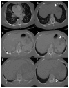
Figure 1
Figure 1
Bilateral mobile thoracolithiasis in a 70-year-old female. Axial CT images show a large right pleural stone (white arrow) and a small left pleural stone (white arrowhead), both of which shifted in position between presentation (A, B), 6-month prior (C, D) and 2-month follow-up (E, F). The larger stone on the right changed positions between the mid-posterior pleural space (A) to lower-posterior pleural space (C, E). The smaller stone on the left moved from the anterior pleural space (B, D) to the posterior pleural space (F). All CT images were acquired on a 64-slice GE LightSpeed VCT scanner. Images A–B were acquired using our routine CT pulmonary angiogram protocol (contrast enhanced acquisition at peak pulmonary opacification with 1.25mm overlapping collimation utilizing 60ml of iodixanol 320 contrast at 120kV with tube current modulation and adaptive statistical iterative reconstruction); images C–D using our routine CT abdomen with contrast protocol (contrast enhanced acquisition at portal venous phase with 3mm overlapping collimation utilizing 80ml of iodixanol 240 contrast at 120kV with tube current modulation and adaptive statistical iterative reconstruction); and images E–F using our routine CT thorax without contrast protocol (contrast enhanced acquisition at arterial phase with 2.5mm overlapping collimation utilizing 60ml of iodixanol 240 contrast at 120kV with tube current modulation and adaptive statistical iterative reconstruction).
Figure 2
Figure 2
Thoracolithiasis in a 70-year-old female. Plain radiographs of the chest (A) and abdomen (B) show a round calcific density located in the right posterior costophrenic angle (white arrows).

References

    1. Kinoshita F, Saida Y, Okajima Y, et al. Thoracolithiasis: 11 cases with a calcified intrapleural loose body. J Thorac Imaging. 2010;25:64–7. - PubMed
    1. Dias AR, Zerbini EJ, Curi N. Pleural stone a case report. J Thorac Cardiovasc Surg. 1968;56:120–2. - PubMed
    1. Puengjesada S, Gupta P, Mottershaw AM. Thoracolithiasis: a case report. Clinical Imaging. 2012:228–30. - PubMed
    1. Kosaka S, Kondo N, Sakaguchi H, et al. Thoracolithiasis. Japanese J Thorac Cardiovasc Surg. 2000;48(5):318–21. - PubMed
    1. Tanaka D, Niwatsukino H, Fujiyoshi F, et al. Thoracolithiasis-a mobile calcified nodule in the intrathoracic space: radiographic, CT and MRI findings. Radiat Med. 2002;20(3):131–3. - PubMed

Publication types