Case Reports
. 2014 Sep 30;8(9):25-9.
doi: 10.3941/jrcr.v8i9.2081. eCollection 2014 Sep.

Posterior rectus sheath hernia causing intermittent small bowel obstruction

Affiliations
Case Reports

Posterior rectus sheath hernia causing intermittent small bowel obstruction

Scott Lenobel et al. J Radiol Case Rep. .

Abstract

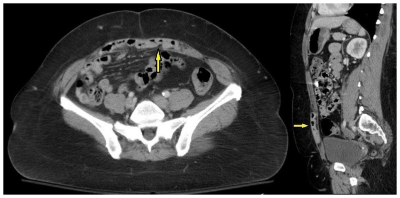
A posterior rectus sheath hernia is an abdominal wall hernia that is rarely encountered. Owing to its rarity, it can be easily overlooked in the setting of a patient presenting with abdominal pain. We report a case of a posterior rectus sheath hernia that caused intermittent small bowel obstruction. The unusual aspects of this case are that the defect was large, measuring 6 cm in the transverse diameter, and that it contained small bowel within a large portion of the rectus sheath. Because the defect was large and affected nearly the entire posterior rectus sheath, it was difficult to discern on computed tomography until a small bowel obstruction developed. In this case, a limited awareness of this clinical entity contributed to the delay in diagnosis.

Keywords: CT; abdomen; abdominal pain; hernia; rectus sheath; small bowel obstruction.


Figures

Figure 1
Figure 1
51 year old female with a posterior rectus sheath hernia. FINDINGS: Supine abdominal radiograph shows a dilated loop of bowel in the upper abdomen (arrow), a nonspecific finding. TECHNIQUE: Supine abdominal radiograph.
Figure 2
Figure 2
51 year old female with a posterior rectus sheath hernia. FINDINGS: Axial multidetector CT of the abdomen (a), sagittal reconstruction (b), and magnified axial view (c) with intravenous and oral contrast show multiple dilated loops of small bowel consistent with a small bowel obstruction. In retrospect, these small bowel loops extend through a large defect in the posterior rectus sheath (arrows). TECHNIQUE: GE Lightspeed VCT. Axial CT with sagittal reconstruction, 654 mA, 120 kV, 5 mm axial mm slices, 3 mm sagittal reconstructions, Oral contrast-Omnipaque 240 50 ml + 850 ml water, IV contrast-Isovue-370 100 ml
Figure 3
Figure 3
51 year old female with a posterior rectus sheath hernia. FINDINGS: Axial multidetector CT of the abdomen (a), sagittal reconstruction (b), and magnified axial view (c) with intravenous contrast only were read as normal. In retrospect, a large posterior rectus sheath defect contains multiple loops of small bowel (arrows) although no small bowel obstruction is present. TECHNIQUE: GE Lightspeed VCT. Axial CT with sagittal reconstruction, 704 mA, 120 kV, 3 mm axial slices, 5 mm sagittal reconstructions, Oral contrast-none, IV contrast-Isovue-370 100 ml.
Figure 4
Figure 4
51 year old female with a posterior rectus sheath hernia. FINDINGS: Axial multidetector CT of the abdomen (a), sagittal reconstruction (b), and magnified axial view (c) with intravenous and oral contrast show multiple dilated loops of small bowel consistent with a small bowel obstruction. The small bowel loops herniate through a large defect in the posterior rectus sheath fascia and abut the anterior abdominal wall (arrows). TECHNIQUE: GE Lightspeed VCT. Axial CT with sagittal reconstruction, 654 mA, 120 kV, 3 mm axial slices, 5 mm sagittal reconstructions, Oral contrast-Omnipaque 240 50 ml + 850 ml water, IV contrast-Isovue-370 100 ml.

References

    1. Losanoff JE, Basson MD, Gruber SA. Spontaneous hernia through the posterior rectus abdominis sheath: case report and review of the published literature 1937–2008. Hernia. 2009;13:555–558. - PubMed
    1. Yang JD, Hwang HP, Kim JH, et al. Development of the rectus abdominis and its sheath in the human fetus. Yonsei Med J. 2012;53:1028–1035. - PMC - PubMed
    1. Felfel M, El Khoury M, Marboeuf Y, Strohl D, Menu Y. Incarcerated hernia through the posterior rectus sheath. AJR Am J Roentgenol. 2005;185:1185–1186. - PubMed
    1. Whitson BA, Ose KJ. Spontaneous posterior rectus sheath hernia: a new clinical entity? Hernia. 2007;11:445–447. - PubMed
    1. Rutkow IM. Demographic and socioeconomic aspects of hernia repair in the United States in 2003. Surg Clin North Am. 2003;83:1045–1051. v–vi. - PubMed

Publication types

MeSH terms