. 2014 Oct 7;5(11):3833-47.
doi: 10.1364/BOE.5.003833. eCollection 2014 Nov 1.

Motion analysis and removal in intensity variation based OCT angiography

Affiliations

Motion analysis and removal in intensity variation based OCT angiography

Xuan Liu et al. Biomed Opt Express. .

Abstract

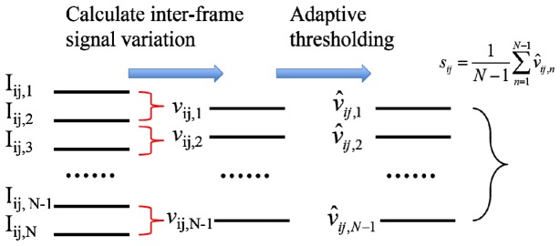
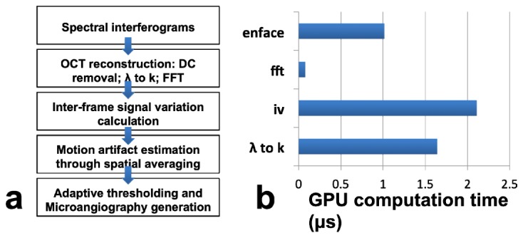
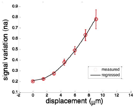
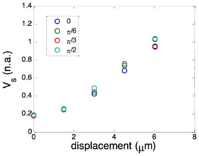
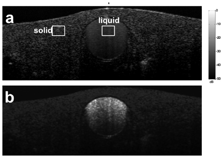
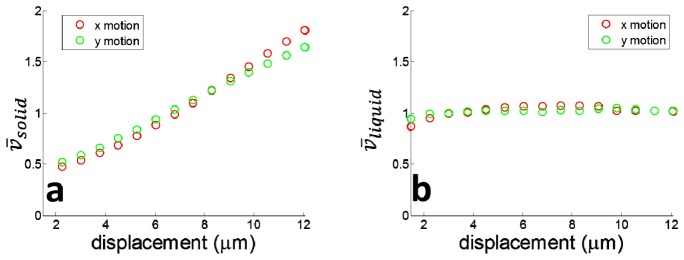
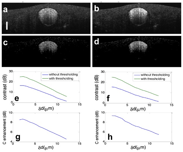
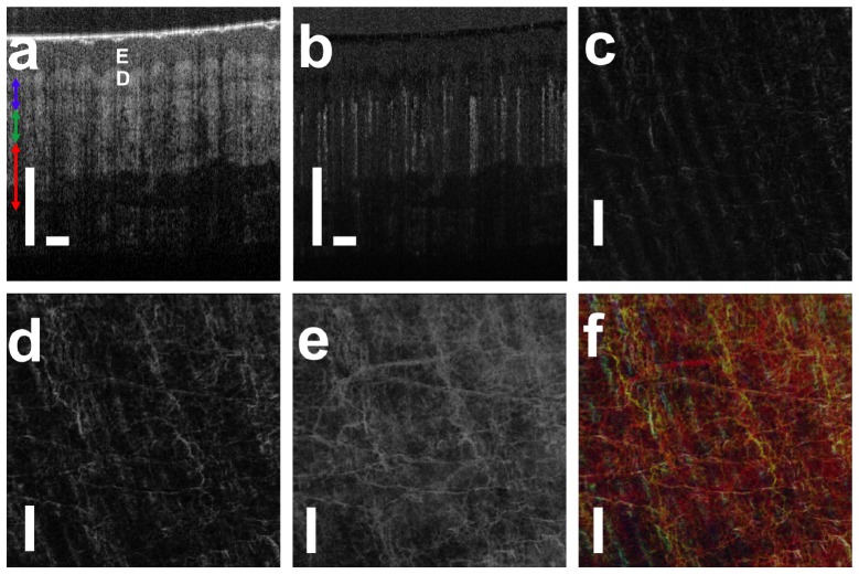
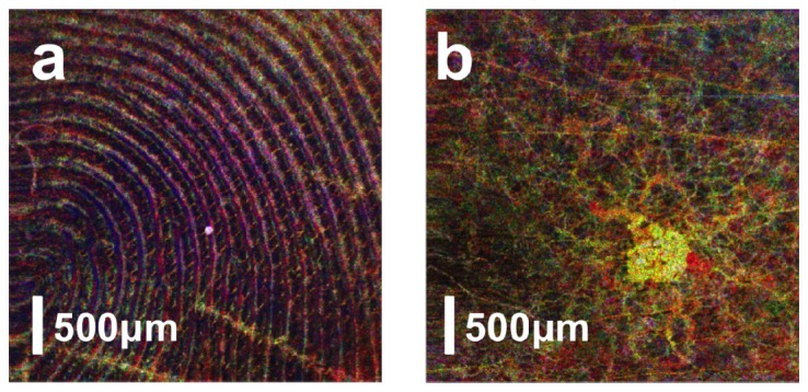
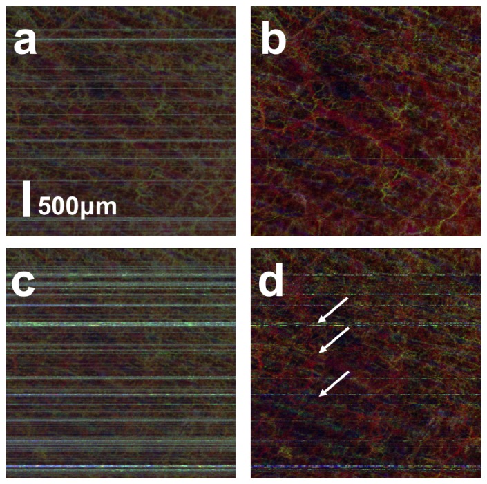
In this work, we investigated how bulk motion degraded the quality of optical coherence tomography (OCT) angiography that was obtained through calculating interframe signal variation, i.e., interframe signal variation based optical coherence angiography (isvOCA). We demonstrated theoretically and experimentally that the spatial average of isvOCA signal had an explicit functional dependency on bulk motion. Our result suggested that the bulk motion could lead to an increased background in angiography image. Based on our motion analysis, we proposed to reduce image artifact induced by transient bulk motion in isvOCA through adaptive thresholding. The motion artifact reduced angiography was demonstrated in a 1.3μm spectral domain OCT system. We implemented signal processing using graphic processing unit for real-time imaging and conducted in vivo microvasculature imaging on human skin. Our results clearly showed that the adaptive thresholding method was highly effective in the motion artifact removal for OCT angiography.

Keywords: (100.2000) Digital image processing; (110.4500) Optical coherence tomography; (170.2655) Functional monitoring and imaging.


Figures

Fig. 1
Fig. 1
Reconstruction of motion artifact reduced OCT angiography.
Fig. 2
Fig. 2
(a) Data processing flowchart of motion artifact reduced angiography; (b) Time expenditure of each processing step preformed on the GPU.
Fig. 3
Fig. 3
Measured spatial average of intensity variation (red circles with errorbar) and fitted curve (black).
Fig. 4
Fig. 4
Spatial average of motion image obtained with BM that has different magnitude and direction.
Fig. 5
Fig. 5
Structural (a) and flow (b) image of a phantom with a polyimide tube filled with bovine milk embedded in solid scattering medium
Fig. 6
Fig. 6
(a) υ¯solid increase as motion in both x and y directions; (b) υ¯liquid remains almost constant with bulk motion
Fig. 7
Fig. 7
Cross-sectional flow image obtained without adaptive thresholding when bulk motion was in x dimension (a) and y dimension (b); Cross-sectional flow image obtained with adaptive thresholding when bulk motion was in x dimension (c) and y dimension (d); contrast of flow image, with (green) and without (blue) adaptive thresholding at different magnitude of motion in x direction (e) and y direction (f); contrast enhancement through adaptive thresholding when motion was in x direction (g) and y direction (h).
Fig. 8
Fig. 8
(a) structural OCT image of human palm skin (E: epidermis; D: dermis); (b) cross-sectional angiography image that highlights blood vessel; (c) – (e) en face microvasculature image in small, medium and large depths; (f) microvasculature image that encodes signal depth with color.
Fig. 9
Fig. 9
isvOCA image obtained from human fingertip (a) and a small skin lesion (b).
Fig. 10
Fig. 10
Angiography image obtained (a) with small magnitude of BM without adaptive thresholding; (b) with small magnitude of BM with adaptive thresholding; (c) with large magnitude of BM without adaptive thresholding; (b) with large magnitude of BM with adaptive thresholding.

References

    1. Chen Z., Milner T. E., Srinivas S., Wang X., Malekafzali A., van Gemert M. J. C., Nelson J. S., “Noninvasive imaging of in vivo blood flow velocity using optical Doppler tomography,” Opt. Lett. 22(14), 1119–1121 (1997).10.1364/OL.22.001119 - DOI - PubMed
    1. Zhao Y., Chen Z., Saxer C., Xiang S., de Boer J. F., Nelson J. S., “Phase-resolved optical coherence tomography and optical Doppler tomography for imaging blood flow in human skin with fast scanning speed and high velocity sensitivity,” Opt. Lett. 25(2), 114–116 (2000).10.1364/OL.25.000114 - DOI - PubMed
    1. Izatt J. A., Kulkarni M. D., Yazdanfar S., Barton J. K., Welch A. J., “In vivo bidirectional color Doppler flow imaging of picoliter blood volumes using optical coherence tomography,” Opt. Lett. 22(18), 1439–1441 (1997).10.1364/OL.22.001439 - DOI - PubMed
    1. Makita S., Hong Y., Yamanari M., Yatagai T., Yasuno Y., “Optical coherence angiography,” Opt. Express 14(17), 7821–7840 (2006).10.1364/OE.14.007821 - DOI - PubMed
    1. Makita S., Fabritius T., Yasuno Y., “Quantitative retinal-blood flow measurement with three-dimensional vessel geometry determination using ultrahigh-resolution Doppler optical coherence angiography,” Opt. Lett. 33(8), 836–838 (2008).10.1364/OL.33.000836 - DOI - PubMed