. 2014 Nov 26;9(11):e113330.
doi: 10.1371/journal.pone.0113330. eCollection 2014.

Initiation of electron transport chain activity in the embryonic heart coincides with the activation of mitochondrial complex 1 and the formation of supercomplexes

Affiliations

Initiation of electron transport chain activity in the embryonic heart coincides with the activation of mitochondrial complex 1 and the formation of supercomplexes

Gisela Beutner et al. PLoS One. .

Abstract

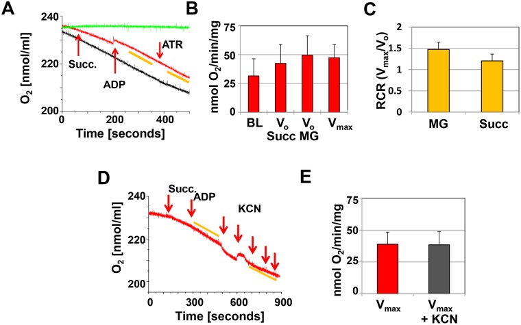
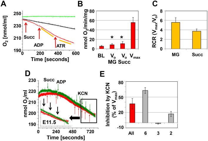
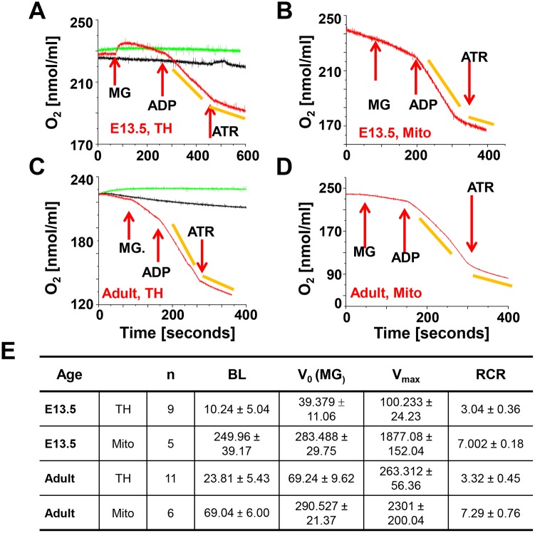
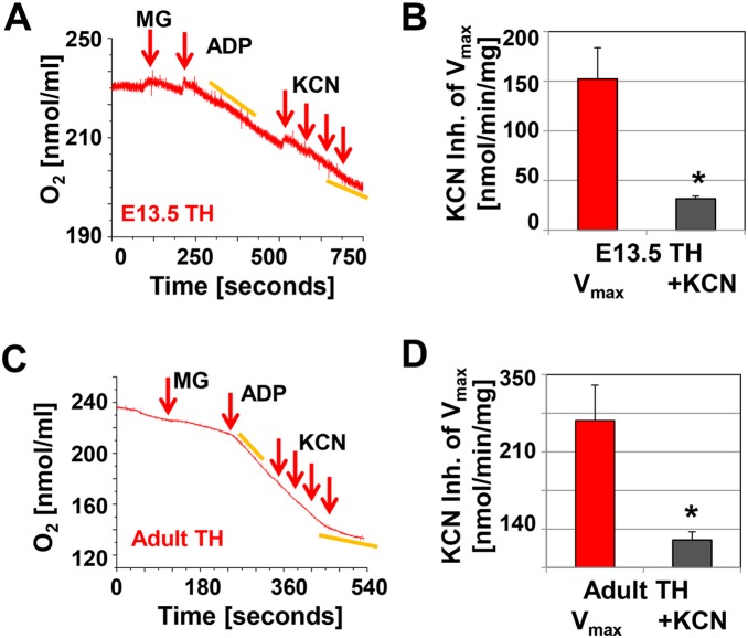
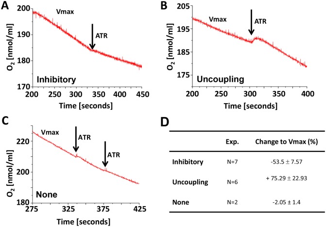
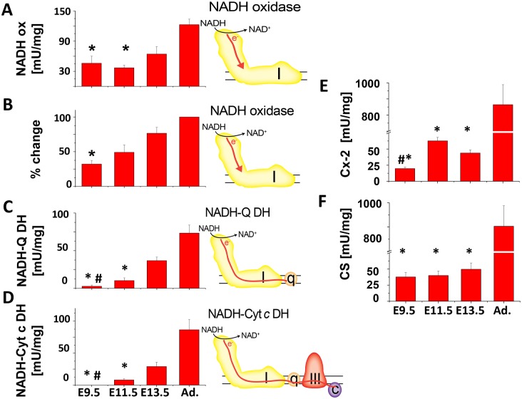
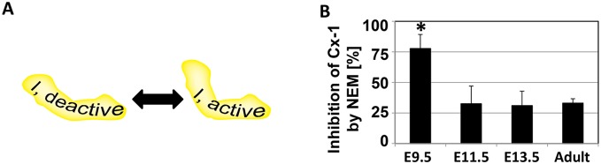
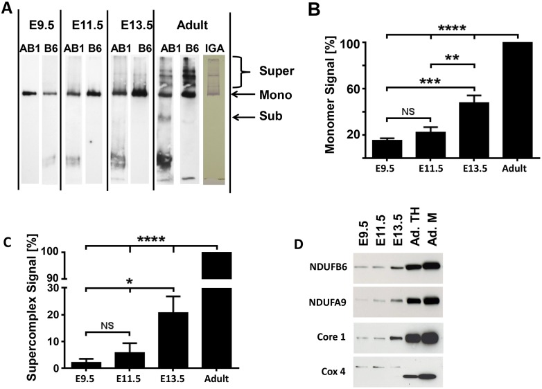
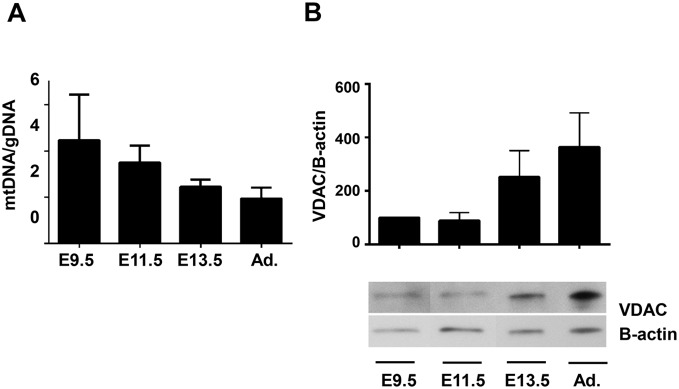
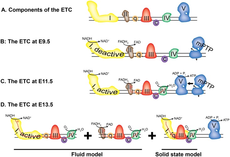
Mitochondria provide energy in form of ATP in eukaryotic cells. However, it is not known when, during embryonic cardiac development, mitochondria become able to fulfill this function. To assess this, we measured mitochondrial oxygen consumption and the activity of the complexes (Cx) 1 and 2 of the electron transport chain (ETC) and used immunoprecipitation to follow the generation of mitochondrial supercomplexes. We show that in the heart of mouse embryos at embryonic day (E) 9.5, mitochondrial ETC activity and oxidative phosphorylation (OXPHOS) are not coupled, even though the complexes are present. We show that Cx-1 of the ETC is able to accept electrons from the Krebs cycle, but enzyme assays that specifically measure electron flow to ubiquinone or Cx-3 show no activity at this early embryonic stage. At E11.5, mitochondria appear functionally more mature; ETC activity and OXPHOS are coupled and respond to ETC inhibitors. In addition, the assembly of highly efficient respiratory supercomplexes containing Cx-1, -3, and -4, ubiquinone, and cytochrome c begins at E11.5, the exact time when Cx-1 becomes functional activated. At E13.5, ETC activity and OXPHOS of embryonic heart mitochondria are indistinguishable from adult mitochondria. In summary, our data suggest that between E9.5 and E11.5 dramatic changes occur in the mitochondria of the embryonic heart, which result in an increase in OXPHOS due to the activation of complex 1 and the formation of supercomplexes.


Conflict of interest statement

Competing Interests: The authors have declared that no competing interests exist.

Figures

Figure 1
Figure 1. Oxygen consumption at E9.5.
A. Representative recording of oxygen consumption in cardiac tissue homogenates (approximately 12.5 µg protein in 0.3 ml respiration buffer). The red recording represents the experiment and the arrows indicate the addition of 2 mM succinate (Succ.), 1 mM ADP and 0.1 mM ATR. The yellow bars indicate the slopes used to calculate oxygen consumption. The black recording represents the tissue homogenate in the respiration medium, but no additions to stimulate oxygen consumption are made. The green recording represents the buffer only. B. Bar graph illustrating oxygen consumption at baseline (BL), after the addition of substrate (Vo Succ and Vo MG) and ADP (Vmax). C. Respiratory control ratio, calculated as Vmax/Vo. A–C: n = 9, for each experiment 7–8 hearts were pooled. D. Representative recording of the inhibition of oxygen consumption by potassium cyanide (KCN) added in 20 µM increments. E. Summary of KCN inhibition (n = 3, 9–10 hearts were pooled per experiment).
Figure 2
Figure 2. Oxygen consumption at E11.5.
A. Representative recording of oxygen consumption in cardiac tissue homogenates (approximately 20 µg protein in 0.3 ml respiration buffer). The red recordings represents the experiment and the arrows indicate the addition of 2 mM succinate (Succ.), 1 mM ADP and 0.1 mM ATR. The yellow bars indicate the slopes used to calculate oxygen consumption. The black recording represents the tissue homogenate in the respiration medium, but no additions to stimulate oxygen consumption are made. The green recording represents the buffer only. B. Bar graph illustrating oxygen consumption at baseline (BL), Vo (malate/glutamate (MG) and Succ), and Vmax. *indicates significance (p≤0.001, ANOVA Vo compared to Vmax). C. Respiratory control ratio, calculated as Vmax/Vo. A–C: n = 14 experiments, for each experiment 2–3 hearts were pooled. D. Representative recordings showing variable inhibition of oxygen consumption by potassium cyanide (KCN) added in 20 µM increments. Red recording: KCN is inhibitory, green recording: no inhibitory effect. E. Summary of KCN inhibition (n = 11; 3–4 hearts were pooled per experiment). All represents the composite data of all experiments, whereas the other columns represent data pooled from experiments with similar results.
Figure 3
Figure 3. Oxygen consumption at E13.5 and in adults.
A and B. Tissue homogenate (TH, approximately 55 µg protein) and isolated mitochondria (Mito) from E13.5 embryos. Hearts of 31 embryos were used to isolate 30 µg of mitochondrial protein for this experiment. C and D. Tissue homogenate (TH, adult) and isolated mitochondria (Mito, adult). 100 µg protein were used per experiment. In all panels the red recordings represents the experiment and the arrows indicate the addition of 3 mM malate and 5 mM glutamate (MG), 1 mM ADP and 0.1 mM ATR. The yellow bars indicate the slopes used to calculate oxygen consumption. The black recording represents the tissue homogenate in the respiration medium, but no additions to stimulate oxygen consumption are made. The green recording represents the buffer only. E. Summary of V0, Vmax and the RCR of E13.5 and adult samples, where n is the number of experiments, BL baseline, MG malate/glutamate, RCR respiratory control ratio. BL, Vo and Vmax are calculated as nmol O2/min/mg.
Figure 4
Figure 4. Inhibition of oxygen consumption by KCN at E13.5 and in adult mice.
A and C. Representative recordings of KCN inhibited oxygen consumption in cardiac tissue homogenates (E13.5 approximately 55 µg protein in 0.3 ml respiration buffer; adult 100 µg protein). The arrows indicate the addition of 3 mM malate and 5 mM glutamate (MG), 1 mM ADP and the addition of KCN (increments of 20 µM). C and D. Bar graphs, showing the inhibition of Vmax in tissue homogenates (TH) by KCN; n = 4, *indicates significance, p≤0.001, T-test.
Figure 5
Figure 5. Variable effect of ATR on mitochondrial respiration at E11.5.
A–C. Representative recordings of the effect of ATR on Vmax; A. inhibitory, B. uncoupling or C. none. D. Summary data of changes to Vmax for each response.
Figure 6
Figure 6. Enzymatic activity of Cx-1, Cx-2 and citrate synthase in cardiac tissue homogenates.
Each assay was done with 1–5 µg protein of cardiac tissue homogenate. The activities are given in mU/mg, except for the dipstick assay, where the relative change was calculated from signal of adult homogenates (100%). In A–D, the cartoon accompanying each graph depicts the flow of electrons through Cx-1 (I) and -3 (III), ubiquinone (q), and cytochrome c (c) tested in the assay. A. NADH-oxidase (NADH ox) assay, *p≤0.05 E9.5 or E11.5 versus adult. B. Cx-1, NADH-oxidase (NADH ox) dipstick assay, *p≤0.05 E9.5 versus adult. C. NADH-ubiquinone dehydrogenase (NADH-Q-DH) assay, *p≤0.05, E 9.5 or E11.5 compared to older embryos or adults; #p≤0.05, E9.5 versus E11.5. D. NADH-cytochrome c dehydrogenase (NADH-Cyt c DH) assay, *p≤0.05, E 9.5 or E11.5 compared to older embryos or adults; #p≤0.05, E9.5 versus E11.5 E. Cx-2/succinate dehydrogenase assay, *p≤0.05, embryos compared to adults; #p≤0.05, E9.5 versus E11.5 and F. Citrate synthase assay. *p≤0.05, embryos compared to adults; #p≤0.05, E9.5 versus E11.5. In all experiments, ANOVA with Tukey post-hoc testing was used and n≥3. All other comparisons were not significant.
Figure 7
Figure 7. At E9.5, Cx-1 is present in its deactive form.
A. The conversion between active and deactive Cx-1 is presumed to be related to structural changes in the multiprotein complex, and oxidation of ND3 leads to permanent deactivation. B. Percent of Cx-1 in deactive state, as measured by the % of NADH oxidase activity that is inhibited by NEM. *p≤0.05 compared to all other ages.
Figure 8
Figure 8. The assembly of ETC complexes into supercomplexes begins around E11.5.
A. One-dimensional high resolution clear native PAGE (hrCN-PAGE) shows the presence of monomeric Cx-1 and the appearance of Cx-1 containing supercomplexes during embryonic development and in adult mouse hearts. Whole tissue homogenates (10 µg for embryonic samples and 5 µg for adult sample) from hearts were solubilized with digitonin (4g digitonin/g protein) and separated by 5–15% hrCN-PAGE. The proteins were then transferred onto nitrocellulose membranes and Cx-1 was visualized first by the detection of NDUFAB1 (AB1, a rabbit polyclonal antibody). Then, the membrane was stripped and re-probed against NDUFB6 (B6, a mouse-monoclonal antibody). In addition, an in-gel assay of Cx-1 (IGA) demonstrates functional complex I monomers and supercomplexes are present in adult heart homogenates. B and C. Quantitative analysis of the presence of monomeric Cx-1 (B) and supercomplexes containing Cx-1 (C). NS–not significant, *p≤0.05, **p≤0.01, ***p≤0.005, and ****p≤0.0001 compared to adult heart samples using ANOVA with Tukey post hoc testing; n = 5. D. Immunocapture of supercomplexes using anti-Cx-3 antibodies; the immunoprecipitate was analyzed by SDS PAGE/immunoblotting for the presence of subunits of Cx-1 (NDUFB6 and NDUFA9), Cx-3 (ubiquinol-cytochrome reductase core protein 1 precursor, Core 1) and Cx-4 (cytochrome c oxidase, subunit 4 (Cox 4)). Homogenates from E9.5, E11.5, and 13.5 heart tissue and homogenate (TH) and mitochondria (M) from adult hearts were examined. Representative immunoblots from n = 3, although these immunoblots are overexposed to demonstrate labeling at younger ages.
Figure 9
Figure 9. Mitochondrial biogenesis and mass measurements.
A. The ratio of mtDNA to gDNA, a measure of mitochondrial biogenesis, was measured in samples of E9.5, 11.5, 13.5 and adult hearts using qPCR to measure levels of CO1 (mtDNA) and 18s (gDNA). B. (Upper) Relative expression of VDAC and beta-actin were evaluated by densitometry and normalized to the value at E9.5 (100%). No significance was determined by ANOVA, n = 4. (Lower) Representative immunoblot demonstrating VDAC and beta-actin expression at each age. Note that lanes, which are not relevant to this manuscript, between E9.5 and E11.5 have been removed.
Figure 10
Figure 10. Model of the embryonic heart ETC.
A. Components of the ETC; the complexes are labeled with Roman numerals. B. At E9.5, the complexes are arranged randomly and do not participate in electron transport, although Cx-1 and -2 have NADH and FADH2 oxidase activity. The mPTP, represented by a not-fully-assembled Cx-5, is open. C. At E11.5, Cx-2, but not Cx-1, participates in electron transport (red line) and oxygen consumption. The mPTP is open or closed. D. At E13.5, the ETC exists in both the fluid (left two panels) and solid (right panel) assembly states, while the mPTP is closed.

References

    1. Porter GA Jr, Hom J, Hoffman D, Quintanilla R, de Mesy Bentley K, et al. Bioenergetics, mitochondria, and cardiac myocyte differentiation. Prog Pediatr Cardiol. 2011;31:75–81. - PMC - PubMed
    1. Ascuitto RJ, Ross-Ascuitto NT. Substrate metabolism in the developing heart. Semin Perinatol. 1996;20:542–563. - PubMed
    1. Lopaschuk GD, Jaswal JS. Energy metabolic phenotype of the cardiomyocyte during development, differentiation, and postnatal maturation. J Cardiovasc Pharmacol. 2010;56:130–140. - PubMed
    1. Baker CN, Ebert SN. Development of aerobic metabolism in utero: Requirement for mitochondrial function during embryonic and fetal periods. OA Biotechnology. 2013;2.
    1. Ingraham CA, Burwell LS, Skalska J, Brookes PS, Howell RL, et al. NDUFS4: creation of a mouse model mimicking a Complex I disorder. Mitochondrion. 2009;9:204–210. - PMC - PubMed

Publication types

MeSH terms

Substances

LinkOut - more resources