. 2014 Dec 16;111(50):E5373-82.
doi: 10.1073/pnas.1408224111. Epub 2014 Nov 26.

The Toll/NF-κB signaling pathway is required for epidermal wound repair in Drosophila

Affiliations

The Toll/NF-κB signaling pathway is required for epidermal wound repair in Drosophila

Lara Carvalho et al. Proc Natl Acad Sci U S A. .

Abstract

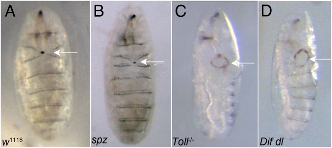
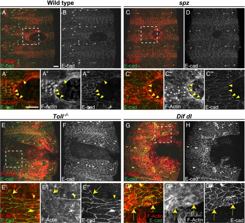
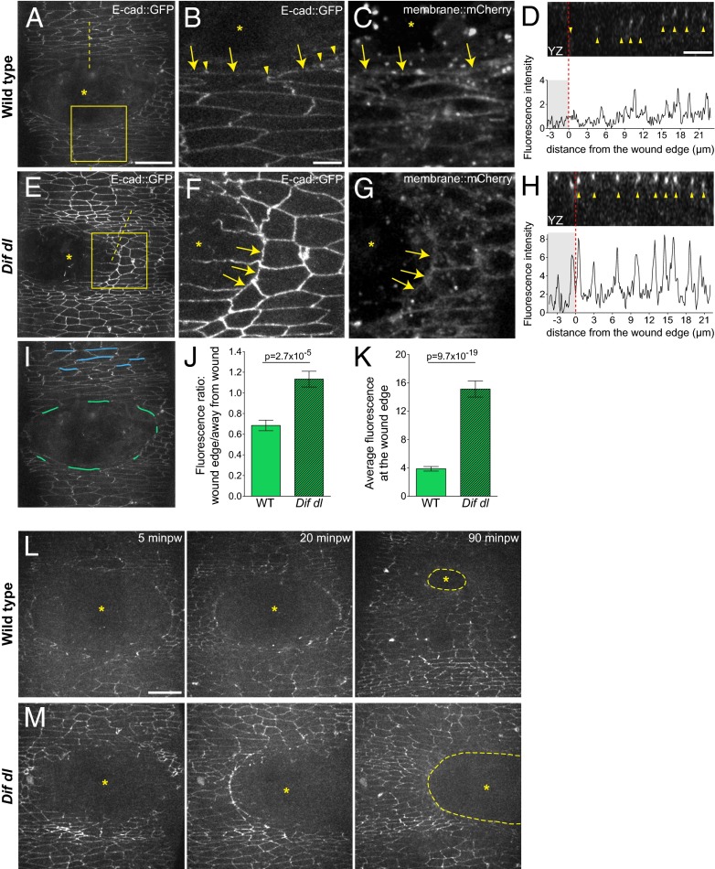
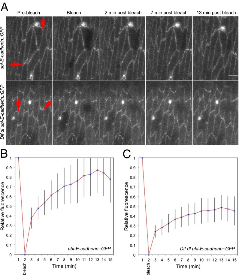
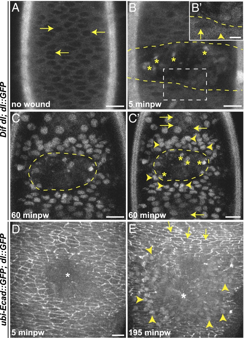
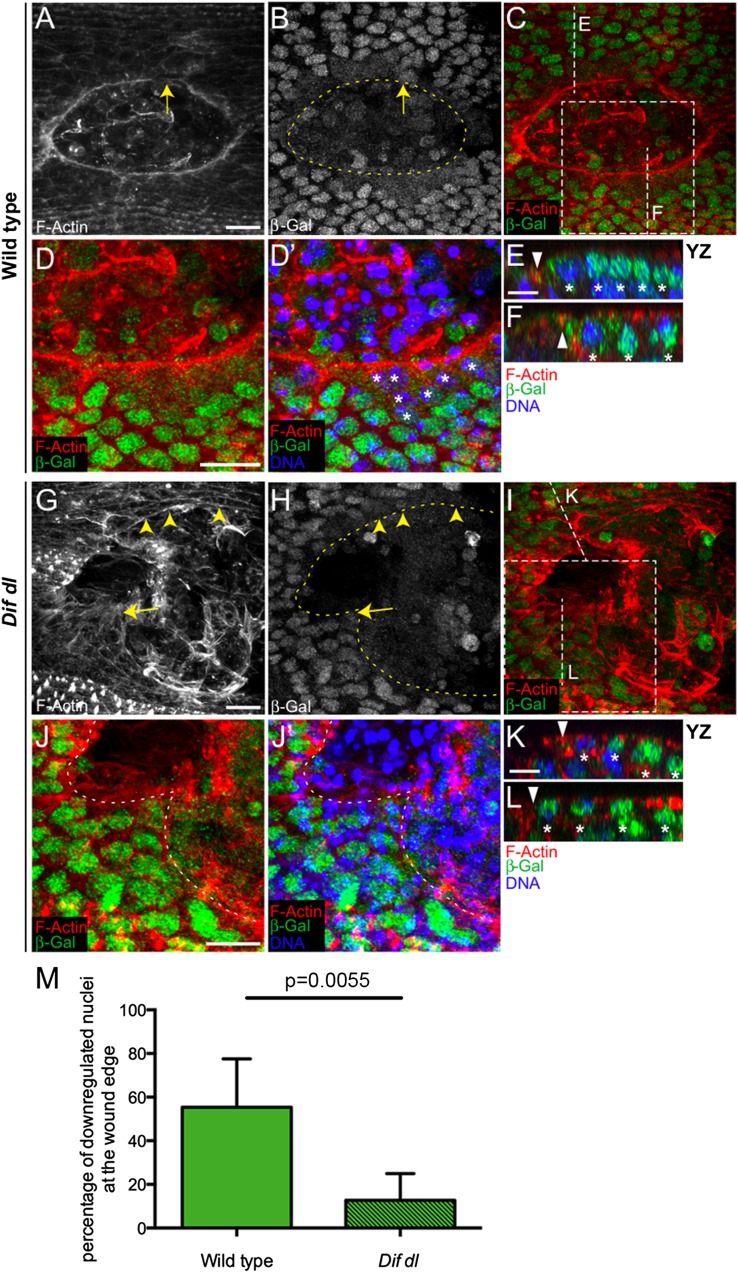
The Toll/NF-κB pathway, first identified in studies of dorsal-ventral polarity in the early Drosophila embryo, is well known for its role in the innate immune response. Here, we reveal that the Toll/NF-κB pathway is essential for wound closure in late Drosophila embryos. Toll mutants and Dif dorsal (NF-κB) double mutants are unable to repair epidermal gaps. Dorsal is activated on wounding, and Dif and Dorsal are required for the sustained down-regulation of E-cadherin, an obligatory component of the adherens junctions (AJs), at the wound edge. This remodeling of the AJs promotes the assembly of an actin-myosin cable at the wound margin; contraction of the actin cable, in turn, closes the wound. In the absence of Toll or Dif and dorsal (dl), both E-cadherin down-regulation and actin-cable formation fail, thus resulting in open epidermal gaps. Given the conservation of the Toll/NF-κB pathway in mammals and the epithelial expression of many components of the pathway, this function in wound healing is likely to be conserved in vertebrates.

Keywords: Drosophila; E-cadherin; NF-κB transcription factors; Toll pathway; epithelial wound repair.


Conflict of interest statement

The authors declare no conflict of interest.

Figures

Fig. 1.
Fig. 1.
Defects in epithelial repair in Toll−/− and Dif dl mutant embryos following laser ablation. (A) Controls (w1118) closed inflicted epidermal lesions in 92.5% of the tested embryos (n = 157). Arrow points to a closed wound. (B) Wounded spz embryos were able to close wounds in the ventral epidermis in 84.2% of the tested mutants (n = 143). Arrow shows a closed wound. (C) Toll−/− embryos failed to close experimentally introduced wounds in 70.3% of the tested mutants (n = 64). Arrow points to an open epidermal gap. (D) Dif dl mutants did not close wounds after laser ablation of a patch of the ventral epidermis in 65.3% of the tested embryos (n = 87). Arrow marks the open wound. Although ablation is always done ventrally, after 16 h of recovery, the open wound could be found on the lateral side in Toll−/− and Dif dl mutants. The number of embryos for each genotype constitutes the combined counts from six to nine independent wounding sessions. The Toll transallelic combination represents a strong loss-of-function phenotype; Dif dl double mutants and spz mutants represent a null phenotype.
Fig. 2.
Fig. 2.
Wounded Toll−/− mutants and Dif dl double mutants do not build a continuous actin cable. Embryos of stage 15 were stained for F-actin (phalloidin) and E-cadherin at 2 h after wounding. (A, C, E, and G) are merged images, whereas (B, D, F, and H) show only E-cadherin. Identical settings were used for all images. Areas for the close-ups are outlined by squares in A, C, E, and G. (A, A′, A′′, A′′′, and B) In wild type, wound-edge cells are contracting and the wound is closing. Wound-edge epidermal cells show a continuous actin cable (A′ and A′′). E-cadherin (B and A′′′, arrowheads) is excluded from the membranes facing the wound. (C, C′, C′′, C′′′, and D) In spz embryos, wound-edge cells are contracting and wound closure is similar to wild type. Wound-edge cells have a continuous actin cable (C′ and C′′). E-cadherin (D and C′′′, arrowheads) is not present at the membranes that face the wound. (E, E′, E′′, E′′′, and F) In Toll−/− embryos, the actin cable is discontinuous. Epidermal cells that maintain high levels of E-cadherin at the membranes bordering the gap (E′′′, arrow) do not assemble an actin cable (E′ and E′′, arrow). In wound-edge cells where some actin bundles are assembled (E′ and E′′, arrowhead), E-cadherin is reduced at the membranes facing the gap (E′′′, arrowhead). (G, G′, G′′, G′′′, and H) In Dif dl embryos, the actin cable is discontinuous. Epidermal cells that maintain high levels of E-cadherin at the membranes bordering the gap (G′′′, arrows) do not assemble an actin cable (G′ and G′′, arrows) at the wound margin. (Scale bars: 10 μm.) Images are the maximum z projections of 11.1- to 22-μm stacks (30–55 slices). Close-ups are the maximum z projections of 5.2- to 18.4-μm stacks (14–46 slices).
Fig. 3.
Fig. 3.
Remodeling of E-cad::GFP at the wound edge fails in Dif dl mutants. (AH) Ventral epidermis of embryos at stage 15 expressing E-cad::GFP and membrane::mCherry. A, B, D–F, and H show E-cad::GFP, whereas C and G show membrane::mCherry. Images are taken with identical settings at 5 min after wounding of wild type (AD) and Dif dl (EH). (B, C, F, and G) are high-magnification images of the wound edge in wild-type and Dif dl embryos (boxed regions in A and E). In wild type, wound-edge membranes have low levels of E-cad::GFP (A; B, arrows) compared with cellular junctions at a distance from the wound. In wound-edge cells, E-cad::GFP is present as apical puncta (B, arrowheads) at contact points between the cells. In Dif dl, all epidermal cells, at the wound edge and far from the wound, have high levels of E-cadherin (E; F, arrows). Wound-edge membranes are outlined by membrane::mCherry (C and G). Asterisks mark the wound. (D and H) YZ cross sections of the epidermis (apical side is up). Dashed lines in A and E mark the positions of the cross-sections. The graphs show the fluorescence intensity profiles of E-cad::GFP in these YZ cross-sections. The wound area is shaded; the wound margin is marked with a red dashed line. Both wild-type and Dif dl embryos display apical localization of E-cad::GFP (arrowheads in D and H). Wild-type cells have low levels of E-cad::GFP at the wound edge, whereas Dif dl cells retain high levels of fluorescence. (I) Schematic representation of the wound-edge membranes (green lines) and AJs (blue lines) used for the measurements and quantification of E-cad::GFP fluorescence shown in J and K. (J) Ratio of E-cad::GFP at wound-edge membranes versus E-cad::GFP at AJs away from the wound in wild type and in Dif dl. In wild type, this ratio is significantly lower (0.685 ± 0.050) than in Dif dl (1.133 ± 0.076), P = 2.7 x 10⁻⁵ (Mann–Whitney test). Error bars are SEM. (K) E-cad::GFP fluorescence intensity at the membranes surrounding the wound in wild type and in Dif dl. Compared with wild type, Dif dl embryos show significantly higher E-cad::GFP fluorescence (P = 9.7 × 10⁻¹⁹, Mann–Whitney test). The value in Dif dl (15.13 ± 1.136) is nearly fourfold higher than in wild type (3.872 ± 0.318). Measurements of 92 wound-edge membranes from 6 wild-type embryos and 91 wound-edge membranes from 5 Dif dl embryos are plotted. Error bars represent SEM. (L) Wound closure dynamics in wild-type embryo expressing E-cad::GFP. Images are single time points from Movie S1. The initial wound of 2,795 μm2 closes rapidly and at 90 min after wounding (minpw), the remaining gap (outlined with a dashed line) is 15-fold smaller: 198.3 μm2. (M) Wound closure dynamics in Dif dl embryo expressing E-cad::GFP. Images are single time points from Movie S3. The initial wound of 2,750 μm2 remains open and at 90 min minpw is approximately 2,157.5 μm2, which is only a 1.3-fold reduction in gap size. (Scale bars: A, E, I, L, and M, 20 μm; BD and FH, 5 μm.)
Fig. 4.
Fig. 4.
E-cad::GFP displays slow rate of FRAP in Dif dl mutants. Cellular interfaces (arrows) in the ventral and ventral-lateral epidermis of embryos at stage 15–16 expressing E-cad::GFP were photobleached. Images were acquired with identical settings. (A, Upper) Time-series images of E-cad::GFP in wild-type embryos (ubi-E-cad::GFP/+). E-cad::GFP recovers rapidly, showing up at the bleached membrane within 2 min after bleaching and reaching approximately 85% recovery at 13 min. (Scale bar: 5 μm.) (A, Lower) Time-series images of E-cad::GFP in Dif dl embryos (Dif dl ubi-E-cad::GFP/Dif dl +). No recovery of E-cad::GFP at the cell membrane was seen in 31.3% of the embryos (n = 16). (Scale bars: 5 μm.) (B) Average fluorescence-recovery kinetics of E-cad::GFP in wild-type embryos (n = 16). (C) Average fluorescence-recovery kinetics of E-cad::GFP in Dif dl embryos (n = 16). E-cad::GFP recovery is impaired in Dif dl cells: At 13 min after bleaching, only approximately 40% of the fusion protein is present at the bleached cellular interface. Error bars represent SD in B and C. The recovery curves are significantly different, P = 0.0005 (Student’s t test).
Fig. 5.
Fig. 5.
Dorsal::GFP undergoes nuclear translocation in epidermal cells on wounding. (AC′) Dorsal::GFP distribution in epidermal cells before and after wounding. Dorsal::GFP is expressed in Dif dl mutant background. Dashed line outlines the wound. Minpw, minutes post wounding. (A) In unwounded embryos, Dorsal::GFP is predominantly cytoplasmic. (B) At 5 minpw, Dorsal::GFP begins to change distribution between cytoplasm and the nuclei. Some cells have less nuclear fluorescence (B′ arrow), whereas in others, the fluorescence is equally distributed between the cytoplasm and the nucleus (B′ arrowhead). (C and C′) At 60 minpw, Dorsal::GFP is exclusively in the nuclei of wound-edge epidermal cells and in several cell rows away from the wound. The activation is graded: arrowheads in C′ show nuclei near the wound with high accumulation of Dorsal::GFP, whereas arrows in C′ point to nuclei that are away from the wound and still have very little Dorsal::GFP. Nuclei that are three to six rows away from the wound display intermediate accumulation of Dorsal::GFP. Asterisks within the wound mark activated Dorsal::GFP in hemocytes. (D and E) Maximum z projections of time-lapse images showing the subcellular localization of Dorsal::GFP and E-cad::GFP in wounded wild-type epidermis. Asterisks mark the wound. Dorsal::GFP and E-cad::GFP are expressed simultaneously in wild-type background. (D) Immediately after wounding, E-cad::GFP decreases at the wound edge; Dorsal::GFP is not observed in the nuclei of epidermal cells. Cytoplasmic Dorsal::GFP is at a more basal level than the apical E-cad::GFP. (E) At 195 min after wounding, Dorsal::GFP has accumulated prominently in several rows of epidermal nuclei around the wound (E arrowheads); E-cad::GFP is decreased in these cells. By contrast, cells that do not show nuclear accumulation of Dorsal::GFP (E arrows) have high E-cad::GFP fluorescence. (Scale bars: 10 μm except B′, 5 μm.)
Fig. 6.
Fig. 6.
Dif and Dorsal are negative regulators of shotgun transcription. (AL) Ventral epidermis of embryos at stage 15 at 1 h after wounding showing F-actin (phalloidin) in red, β-Gal in green, and DNA (DAPI) in blue. β-Gal stains nuclei with active shotgun transcription because all embryos are heterozygous for P{lacW}shgk03401. Images are the maximum intensity z projections of wild-type (A, B, C, D, and D′) and Dif dl embryos (GJ′) and represent 7.5- to 13-μm stacks (15–26 slices). D, D′, J, and J′ represent close-ups of the boxed areas in C and I, respectively. Identical settings were used for all images. In wild type, a continuous actin cable outlines the smooth wound margin (A). In Dif dl, the wound edge is irregular and the actin cable is discontinuous (G), with regions showing some actin bundles (arrowheads) and regions showing no actin bundles (arrow). In wild type, the nuclear β-Gal in wound-edge cells is greatly decreased compared with nuclei away from the wound (BD′). In Dif dl, many nuclei at the wound margin maintain high β-Gal expression (HJ′). Nuclei with high levels of β-Gal belong to cells where the actin cable is missing (arrow in G and H). Dashed lines in B, H, J, and J′ outline the wound edge. (E, F, K, and L) YZ slices of the regions shown by vertical lines in C and I. Triangles mark the wound edge/actin cable and asterisks mark the epidermal nuclei. The wound is to the left. In wild type, nuclei at the wound edge have a decreased β-Gal compared with nuclei away from the edge (E and F). In Dif dl, β-Gal fluorescence is similar in edge and nonedge nuclei where the actin cable is missing (L), but down-regulated in wound-edge nuclei where some actin cable is present (K). (M) Percentage of cells at the wound edge with decreased nuclear β-Gal at 1 h after injury. In wild type, 55.4 ± 22.2% of the wound-edge cells have decreased nuclear β-Gal, whereas in Dif dl, 12.7 ± 12.3% of the wound-edge cells show β-Gal down-regulation; P = 0.0055 (Student’s t test). Error bars are SD. Five wild-type and five Dif dl embryos were quantified. All wound-edge nuclei were measured as follows: 100 wild-type and 83 Dif dl. (Scale bars: AD′ and GJ′, 10 μm; E, F, K, and L, 5 μm.)

References

    1. Martin P, Lewis J. Actin cables and epidermal movement in embryonic wound healing. Nature. 1992;360(6400):179–183. - PubMed
    1. Kiehart DP, Galbraith CG, Edwards KA, Rickoll WL, Montague RA. Multiple forces contribute to cell sheet morphogenesis for dorsal closure in Drosophila. J Cell Biol. 2000;149(2):471–490. - PMC - PubMed
    1. Garcia-Fernandez B, Campos I, Geiger J, Santos AC, Jacinto A. Epithelial resealing. Int J Dev Biol. 2009;53(8–10):1549–1556. - PubMed
    1. Schäfer M, Werner S. Transcriptional control of wound repair. Annu Rev Cell Dev Biol. 2007;23:69–92. - PubMed
    1. Mace KA, Pearson JC, McGinnis W. An epidermal barrier wound repair pathway in Drosophila is mediated by grainy head. Science. 2005;308(5720):381–385. - PubMed

Publication types

MeSH terms