. 2014 Dec 16;42(22):13824-38.
doi: 10.1093/nar/gku1171. Epub 2014 Nov 26.

Sequential packaging of RNA genomic segments during the assembly of Bluetongue virus

Affiliations

Sequential packaging of RNA genomic segments during the assembly of Bluetongue virus

Po-Yu Sung et al. Nucleic Acids Res. .

Abstract

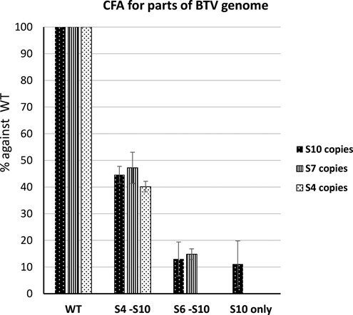
Bluetongue virus (BTV), a member of the Orbivirus genus within the Reoviridae family, has a genome of 10 double-stranded RNA segments, with three distinct size classes. Although the packaging of the viral genome is evidently highly specific such that every virus particle contains a set of 10 RNA segments, the order and mechanism of packaging are not understood. In this study we have combined the use of a cell-free in vitro assembly system with a novel RNA-RNA interaction assay to investigate the mechanism of single-stranded (ss) RNAs packaging during nascent capsid assembly. Exclusion of single or multiple ssRNA segments in the packaging reaction or their addition in different order significantly altered the outcome and suggested a particular role for the smallest segment, S10. Our data suggests that genome packaging probably initiates with the smallest segment which triggers RNA-RNA interaction with other smaller segments forming a complex network. Subsequently, the medium to larger size ssRNAs are recruited until the complete genome is packaging into the capsid. The untranslated regions of the smallest RNA segment, S10, is critical for the instigation of this process. We suggest that the selective packaging observed in BTV may also apply to other members of the Reoviridae family.


Figures

Figure 1.
Figure 1.
Exclusion of specific BTV RNA segment influences genome packaging. An incomplete set of 32P-labelled BTV ssRNAs that excludes S2 or S5 or S10 (indicated as -S2, -S5, -S10) but includes all the 9 respective segments were used in the in vitro CFA assay; the reaction mixture was purified on a sucrose gradient. A complete set of 10 ssRNAs was also included (all 10) as a control. (A) RNA distribution within the sucrose gradient fractions 2–9 was analysed on an agarose gel. The fraction containing assembled cores (fraction 6) is indicated with an asterix (*). (B) Packaged RNA profile after RNAse digestion and purification of fraction 6 analysed by 1% denaturing agarose gel. Segments S1–S10 are indicated on the left. (C) Quantification of the effect of segment exclusion. Ten BTV ssRNAs (WT) or ssRNAs excluding one ssRNA at a time (-S1, -S2, etc.) were used in the CFA assay. Packaged ssRNA in relevant fraction containing cores was purified on a sucrose gradient. BTV segment S6 was quantified by qRT-PCR to represent the packaging efficiency. Quantities of S6 in samples of -S1, -S2, etc. were compared with WT control in the same experiment and packaging efficiency was calculated. Standard deviations from three independent experiments are indicated (error bars).
Figure 2.
Figure 2.
Exchanging UTRs of segment S10 influences packaging in vitro. (A) Schematic representation of chimeric segments S10. The UTRs of BTV-1 S10 (white boxes), flanking the open reading frame (grey boxes) were replaced with UTRs (pattern boxes) of BTV-1 S3 (S3/S10) or S5 (S5/S10) or S8 (S8/S10) or BTV-10 S10 (B10/B1) or AHSV-4 S10 (A4/B1) using overlap PCR and T7 transcription. (B) BTV-1 S10 (WT) or chimeric S10 (S3/S10, or S5/S10, or S8/S10, or B10/B1, or A4/B1) were included with segments S1–S9 in CFA assay. Packaged RNAs were purified and the encapsidation of S10 in each assay was measured by qRT-PCR using specific primers for the BTV-1 S10 coding region. The packaging efficiency and standard deviations (error bars) for each condition was calculated and normalized considering WT conditions as 100%.
Figure 3.
Figure 3.
Effect of exchanging UTRs of S10 in packaging using an in vivo system. (A) A cartoon shows the process of in vivo single replication packaging assay. A modified sequence that can be specifically detected and quantified by PCR (marked with *) was introduced in the chimeric S10 ssRNAs used for transfection (in dark grey). Note that 12–16 h after transfection and infection, newly formed cores were purified and the amount of modified RNA packaged within the cores was measured. (B) Quantification of modified S10 (WT, S3/S10, S5/S10, S8/S10, B10/B1 and A4/B1) packaged in the new viral cores was correlated with the total quantity of new cores in the sample to obtain the packaging efficiency. The data was standardized to the wild-type data considered to be 100% and the ratios were calculated. Standard deviations are indicated as error bars.
Figure 4.
Figure 4.
Effects of chimeric S10 on virus recovery using RG system. A complete set of 9 BTV-1 segments (S1–S9) and one of the chimeric S10 (S3/S10 to A4/B1) or the WT S10 were used in RG system. (A) Recovered viruses were amplified once and analysed by plaque assay on monolayers of BSR cells. (B) Genomic dsRNA of reassortant virus containing BTV-10/BTV-1 chimeric S10 (B10/B1) was purified and the sequence was confirmed by RT-PCR and sequencing. Nucleotides specific to BTV-10 in the electropherogram and in the actual sequence are indicated with an asterix. The sequencing data shows the reverse complement strand. (C) Deletion in UTRs suppresses packaging. BTV-1 S10 (WT), 5′ UTR and both UTRs truncated S10 (Δ5UTR and ΔUTRs) and a series of 3′-end truncated S10 (Δ12, Δ35, Δ60) were tested using CFA assay as described in Figure 2B.
Figure 5.
Figure 5.
Complete set of BTV RNA segments is required for RNA packaging. Full set of BTV-1 10 ssRNAs (WT), S4–S10 (S4-S10), S6–S10 (S6-S10) or S10 only were used in CFA system to determine the packaging efficiency as described in Figures 1 and 2. Three segments (S4, S7 and S10; shown in different patterned bars as indicated) were detected with qRT-PCR when applicable. The packaging efficiencies are shown in percentage and standard deviation (error bars) were calculated.
Figure 6.
Figure 6.
A schematic for RNA–RNA interaction assay based on BTV S10-coated beads.
Figure 7.
Figure 7.
BTV S10 interacts with smaller segments. (A) Beads coated with a S10-specific primer were incubated sequentially with BTV-1 S10 and with 1 pmol of segments S1–S9 individually. Interaction with RRV S9 was included as a negative control. After extensive washing, the attached RNA was released by heating. The amount of interacting RNA was determined by qRT-PCR using primers specific to each segment. The copy number was correlated by minus non-specific binding detected in beads-only control. S10 and the standard deviations from three individual experiments are indicated. (B) Beads coated with (+) or without (−) BTV-1 S10 were similarly prepared and sequentially incubated with 32P-labelled S1, S3, S6 or S8. After three washes, RNAs were heat-released and analysed on a denaturing agarose gel and Phospho-imager exposure. The black arrows indicate positive interaction. (C) The interaction between S8 and truncated S10 was measured with a similar method: beads were coated with BTV-1 S10 (S10), S10 lacking 5′ and 3′ UTRs (ΔUTRs), 3′ UTR (Δ3′UTR) or 5′ UTR (Δ5′UTR) and incubated with equal amounts of BTV-1 S8. Interacting S8 was analysed and quantified similarly. Interaction rates and standard deviation (error bars) were calculated.
Figure 8.
Figure 8.
Smaller segments link larger segments to S10 in a specific order. S10-coated beads were prepared as described in Figure 6. (A) 1 pmol BTV-1 S1, S5 or RRV S9 were incubated with S10 beads only (S10) or together with a mixture of S6, S7, S8 and S9 (S6-S10). Interacting ssRNAs were analysed by qRT-PCR and correlated to the control lacking S10. Standard deviations from three individual experiments are indicated (error bars). (B) Enhancement of the interaction between S5 and S10. BTV-1 S5 was incubated with S10 beads alone (S10) or with S6, S7, S8 or S9 separately (S10 + S6, etc.) or with a mixture of S7 to S9 (S10 + S7-S9) or S6 to S9 (S10 + S6-S9). (C) The interaction between S1 and S10 was enhanced when adding a different mixture of segments. BTV-1 S1 was incubated with S10 beads alone (S10) or with a mixture containing S6 to S9 (S6-S10), or S4 to S9 (S4-S10), or S2 to S9 (S2-S10).
Figure 9.
Figure 9.
Specific interaction order among segments with different sizes. (A) Smaller segments link larger segment S1–S8. S8-coated beads were prepared similarly as S10-coated beads as described. BTV-1 S1 was incubated with S8 beads alone (S8) or with mixtures of S8 to S10 (S8-S10), or S4 to S10 (S4-S10), or S2 to S10 (S2-S10). The pulled-down S1 was quantified with qRT-PCR as described. (B) Larger segments also link smaller segment S10 to S3. S3-coated beads were similarly prepared. BTV-1 S10 was incubated with S3 beads alone (S10) or with different mixtures (S1-S3, or S1-S6, or S1-S9). (C) S3 beads were incubated with BTV-1 S1 and S2 or with all other 9 segments. The pulled-down S1 and S2 were quantified as described. The increased interaction was shown in bar and standard deviation (error bars) were calculated.

References

    1. Roy P. Bluetongue virus: dissection of the polymerase complex. J. Gen. Virol. 2008;89:1789–1804. - PMC - PubMed
    1. Mertens P.P., Brown F., Sangar D.V. Assignment of the genome segments of bluetongue virus type 1 to the proteins which they encode. Virology. 1984;135:207–217. - PubMed
    1. Roy P. Functional mapping of bluetongue virus proteins and their interactions with host proteins during virus replication. Cell Biochem. Biophys. 2008;50:143–157. - PubMed
    1. Roy P., Purdy M.A., Petre J., Rao C.D. Cloning and nucleotide sequencing of bluetongue virus genomes. Prog. Clin. Biol. Res. 1985;178:363–370. - PubMed
    1. Verwoerd D.W., Els H.J., De Villiers E.M., Huismans H. Structure of the bluetongue virus capsid. J. Virol. 1972;10:783–794. - PMC - PubMed

Publication types