. 2015 Jan;43(Database issue):D345-56.
doi: 10.1093/nar/gku1214. Epub 2014 Nov 26.

The RCSB Protein Data Bank: views of structural biology for basic and applied research and education

Affiliations

The RCSB Protein Data Bank: views of structural biology for basic and applied research and education

Peter W Rose et al. Nucleic Acids Res. 2015 Jan.

Abstract

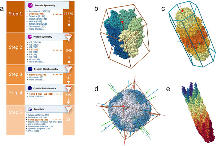
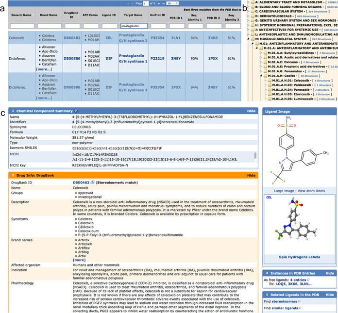
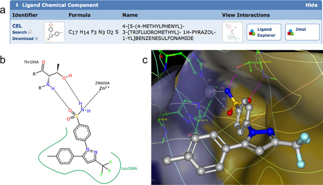
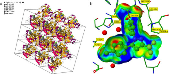
The RCSB Protein Data Bank (RCSB PDB, http://www.rcsb.org) provides access to 3D structures of biological macromolecules and is one of the leading resources in biology and biomedicine worldwide. Our efforts over the past 2 years focused on enabling a deeper understanding of structural biology and providing new structural views of biology that support both basic and applied research and education. Herein, we describe recently introduced data annotations including integration with external biological resources, such as gene and drug databases, new visualization tools and improved support for the mobile web. We also describe access to data files, web services and open access software components to enable software developers to more effectively mine the PDB archive and related annotations. Our efforts are aimed at expanding the role of 3D structure in understanding biology and medicine.


Figures

Figure 1.
Figure 1.
(a) Successive drill-down to find protein complexes with C5 point group that are homo-pentamers from human. The drill-down feature can be accessed from the Search panel on the home page. (b) Visualization of a symmetric protein (PDB ID 3EAM) (28) with a 5-fold axis (red), a radial color gradient to emphasize rotational symmetry and a pentagonal prism. (c) Dihedral (D7) complex (PDB ID 1YA7) (29), composed of three types of subunits (different colors). (d) Octahedral complex (PDB ID 1SHS) (30) with 4-fold (red), 3-fold (green) and 2-fold (green) axes, surrounded by an octagon. (e) Helical complex (PDB ID 1IFD) (31), displayed with a color gradient to emphasize the helical pattern of subunits.
Figure 2.
Figure 2.
(a) Section of drug mapping table for prostaglandins with some of the hits shown for Celexocib and Diclofenac. On the right side of the table are links to related PDB entries and% sequence identity compared to the target sequence. (b) ATC tree with anti-inflammatory section expanded, showing Celecoxib and related drugs represented in the PDB. (c) Ligand summary page for Celecoxib, enhanced with DrugBank annotation (orange box). At the bottom right are links to all four entries that contain Celecoxib, plus a search link to find similar ligands.
Figure 3.
Figure 3.
(a) Ligand Chemical Component widget on the Structure Summary page for Celecoxib bound to carbonic anhydrase II from PDB ID 1OQ5 (41). The ‘View Interactions’ section provides access to 2D and 3D ligand-binding site views. (b) 2D Ligand interaction diagram generated by PoseView (42) (dashed lines, hydrogen bonds and metal interactions; green line, hydrophobic interactions). (c) Interactive 3D visualization using Ligand Explorer (33) (ligand in stick and ball style, and some of the interacting binding site residues in green; dashed lines, hydrogen bonds and metal coordination with distance, and transparent binding site surface color-coded by hydrophobicity of the closest amino acid residue (yellow, hydrophobic; blue, hydrophilic).
Figure 4.
Figure 4.
Two new custom rendering options on the Jmol /JSmol page demonstrated using PDB ID 1OQ5 (Celecoxib bound to Carbonic Anhydrase II) (41). (a) Rendering of the crystal packing (3 × 3 unit cells in the ab plane), carbonic anhydrase II (cartoon style) and Celecoxib (CPK style). (b) Jmol rendering of the Celecoxib (ball-and-stick style) in carbonic anhydrase II binding site (stick style). The trimmed ligand VDW surface is color-coded by the distance of the surface to the nearest side chain atom (short contact distances, red; optimal hydrophobic interaction distances, green; longer distances, blue). The red regions (close contacts) at the top of the surface represent hydrogen bonds and metal coordination of the sulfonamide group of Celecoxib with THR 199 and Zinc.
Figure 5.
Figure 5.
Protein feature view for Calmodulin. The main reference (gray track) is the full-length sequence from UniProt. The top of the view (green) provides a summary of important functional motifs plus UniProt sites. By moving the mouse over any of these regions, more information about this region can be viewed. This is followed by annotations from Pfam (yellow), SCOP domain annotations (orange), computationally inferred information, such as protein disorder score or hydropathy. At the bottom, in blue, are the data derived from PDB. A secondary-structure track shows helical and strand regions along the protein. In the ‘condensed’ (default) view representative protein chains are being displayed mapped to the protein sequence. To view the mapping of all available PDB chains the ‘expanded’ view can be called by pressing the ‘+’ button.
Figure 6.
Figure 6.
Gene view of human chromosome 16, zoomed into the region of the HBA1, hemoglobin α1 gene. The top of the view is a ruler that shows the current region along a chromosome. The blue/red/green/black colored tracks is the color-coding of the nucleotides. The PDB track shows two PDB chains: PDB ID 2W72 chain A (50), PDB ID 1C7C chain A (51) and how they align to this region on the chromosome. The Genes track shows four transcripts, one of which is the main HBA1 coding sequence. By zooming out, the human α globin gene cluster, spanning about 30 kb and including several hemoglobin genes, can be visualized.

References

    1. Berman H.M., Westbrook J., Feng Z., Gilliland G., Bhat T.N., Weissig H., Shindyalov I.N., Bourne P.E. The Protein Data Bank. Nucleic Acids Res. 2000;28:235–242. - PMC - PubMed
    1. Rose P.W., Bi C., Bluhm W.F., Christie C.H., Dimitropoulos D., Dutta S., Green R.K., Goodsell D.S., Prlić A., Quesada M., et al. The RCSB Protein Data Bank: new resources for research and education. Nucleic Acids Res. 2013;41:D475–D482. - PMC - PubMed
    1. Protein Data Bank. Crystallography: Protein Data Bank. Nat. New Biol. 1971;233:223. - PubMed
    1. Berman H.M., Kleywegt G.J., Nakamura H., Markley J.L. The Protein Data Bank at 40: reflecting on the past to prepare for the future. Structure. 2012;20:391–396. - PMC - PubMed
    1. Berman H., Henrick K., Nakamura H. Announcing the worldwide Protein Data Bank. Nat. Struct. Biol. 2003;10:980. - PubMed

Publication types