

Biofilm formation by virulent and non-virulent strains of Haemophilus parasuis
- PMID: 25428823
- PMCID: PMC4245831
- DOI: 10.1186/s13567-014-0104-9
Biofilm formation by virulent and non-virulent strains of Haemophilus parasuis
Abstract
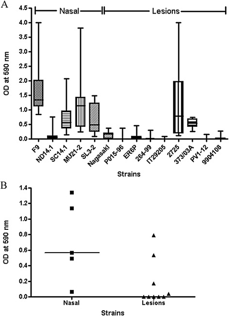
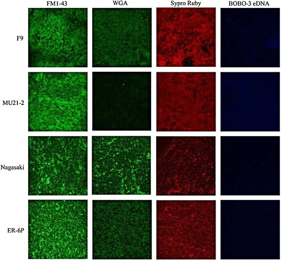
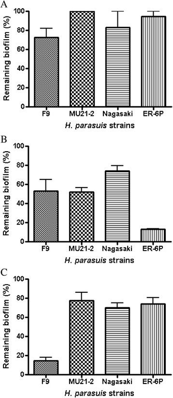
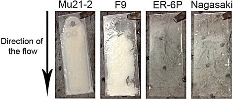
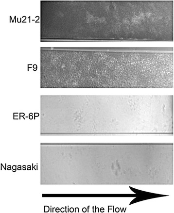
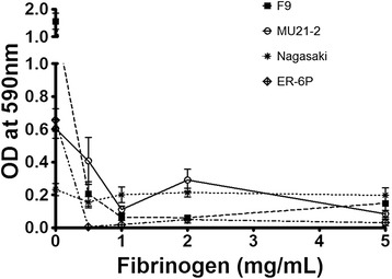
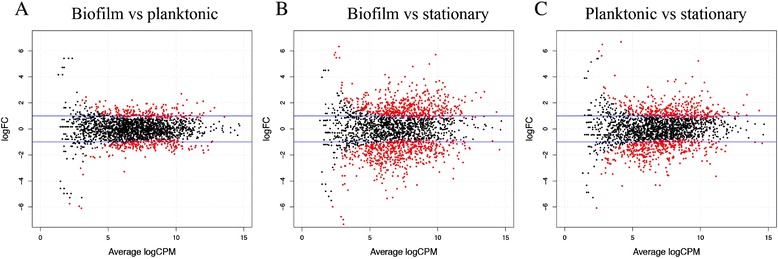
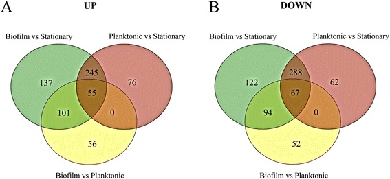
Haemophilus parasuis is a commensal bacterium of the upper respiratory tract of healthy pigs. It is also the etiological agent of Glässer's disease, a systemic disease characterized by polyarthritis, fibrinous polyserositis and meningitis, which causes high morbidity and mortality in piglets. The aim of this study was to evaluate biofilm formation by well-characterized virulent and non-virulent strains of H. parasuis. We observed that non-virulent strains isolated from the nasal cavities of healthy pigs formed significantly (p < 0.05) more biofilms than virulent strains isolated from lesions of pigs with Glässer's disease. These differences were observed when biofilms were formed in microtiter plates under static conditions or formed in the presence of shear force in a drip-flow apparatus or a microfluidic system. Confocal laser scanning microscopy using different fluorescent probes on a representative subset of strains indicated that the biofilm matrix contains poly-N-acetylglucosamine, proteins and eDNA. The biofilm matrix was highly sensitive to degradation by proteinase K. Comparison of transcriptional profiles of biofilm and planktonic cells of the non-virulent H. parasuis F9 strain revealed a significant number of up-regulated membrane-related genes in biofilms, and genes previously identified in Actinobacillus pleuropneumoniae biofilms. Our data indicate that non-virulent strains of H. parasuis have the ability to form robust biofilms in contrast to virulent, systemic strains. Biofilm formation might therefore allow the non-virulent strains to colonize and persist in the upper respiratory tract of pigs. Conversely, the planktonic state of the virulent strains might allow them to disseminate within the host.
Figures

Similar articles
-
Biofilm formation by field isolates and reference strains of Haemophilus parasuis.Vet Microbiol. 2006 Nov 26;118(1-2):117-23. doi: 10.1016/j.vetmic.2006.07.009. Epub 2006 Sep 7. Vet Microbiol. 2006. PMID: 16959443
-
Time course Haemophilus parasuis infection reveals pathological differences between virulent and non-virulent strains in the respiratory tract.Vet Microbiol. 2014 Jun 4;170(3-4):430-7. doi: 10.1016/j.vetmic.2014.01.011. Epub 2014 Feb 3. Vet Microbiol. 2014. PMID: 24613292
-
Advances in the quest for virulence factors of Haemophilus parasuis.Vet J. 2013 Dec;198(3):571-6. doi: 10.1016/j.tvjl.2013.08.027. Epub 2013 Sep 4. Vet J. 2013. PMID: 24084037 Review.
-
Virulence and draft genome sequence overview of multiple strains of the swine pathogen Haemophilus parasuis.PLoS One. 2014 Aug 19;9(8):e103787. doi: 10.1371/journal.pone.0103787. eCollection 2014. PLoS One. 2014. PMID: 25137096 Free PMC article.
-
Haemophilus parasuis: new trends on diagnosis, epidemiology and control.Vet Microbiol. 2004 Mar 26;99(1):1-12. doi: 10.1016/j.vetmic.2003.12.001. Vet Microbiol. 2004. PMID: 15019107 Review.
Cited by
-
One Health-One Biofilm.Vet Res. 2022 Jul 7;53(1):51. doi: 10.1186/s13567-022-01067-4. Vet Res. 2022. PMID: 35799278 Free PMC article.
-
Metatranscriptomics reveals metabolic adaptation and induction of virulence factors by Haemophilus parasuis during lung infection.Vet Res. 2015 Sep 23;46(1):102. doi: 10.1186/s13567-015-0225-9. Vet Res. 2015. PMID: 26395877 Free PMC article.
-
Genetic variants and phylogenetic analysis of Haemophilus parasuis (HPS) OMPP2 detected in Sichuan, China from 2013 to 2015.J Vet Med Sci. 2017 Oct 7;79(10):1648-1651. doi: 10.1292/jvms.16-0519. Epub 2017 Aug 18. J Vet Med Sci. 2017. PMID: 28824043 Free PMC article.
-
The Bordetella Bps Polysaccharide Is Required for Biofilm Formation and Enhances Survival in the Lower Respiratory Tract of Swine.Infect Immun. 2017 Jul 19;85(8):e00261-17. doi: 10.1128/IAI.00261-17. Print 2017 Aug. Infect Immun. 2017. PMID: 28559403 Free PMC article.
-
Strain-dependent interactions of Streptococcus suis and Glaesserella parasuis in co-culture.Can J Vet Res. 2023 Oct;87(4):245-253. Can J Vet Res. 2023. PMID: 37790267 Free PMC article.
References
-
- Aragon V, Segalés J, Oliveira S. Glässer’s disease. In: Zimmerman JJ, Karriker LA, Ramirez A, Schwartz KJ, Stevenson GW, editors. Diseases of Swine. 10. Chichester, UK: Wiley-Blackwell; 2012. pp. 760–769.
Publication types
MeSH terms
Associated data
- Actions
Grants and funding
LinkOut - more resources
Full Text Sources
Other Literature Sources
Medical
Molecular Biology Databases
Miscellaneous
