. 2015 Feb;89(3):1523-36.
doi: 10.1128/JVI.02596-14. Epub 2014 Nov 26.

The nsp3 macrodomain promotes virulence in mice with coronavirus-induced encephalitis

Affiliations

The nsp3 macrodomain promotes virulence in mice with coronavirus-induced encephalitis

Anthony R Fehr et al. J Virol. 2015 Feb.

Abstract

All coronaviruses encode a macrodomain containing ADP-ribose-1"-phosphatase (ADRP) activity within the N terminus of nonstructural protein 3 (nsp3). Previous work showed that mouse hepatitis virus strain A59 (MHV-A59) with a mutated catalytic site (N1348A) replicated similarly to wild-type virus but was unable to cause acute hepatitis in mice. To determine whether this attenuated phenotype is applicable to multiple disease models, we mutated the catalytic residue in the JHM strain of MHV (JHMV), which causes acute and chronic encephalomyelitis, using a newly developed bacterial artificial chromosome (BAC)-based MHV reverse genetics system. Infection of mice with the macrodomain catalytic point mutant virus (N1347A) resulted in reductions in lethality, weight loss, viral titers, proinflammatory cytokine and chemokine expression, and immune cell infiltration in the brain compared to mice infected with wild-type virus. Specifically, macrophages were most affected, with approximately 2.5-fold fewer macrophages at day 5 postinfection in N1347A-infected brains. Tumor necrosis factor (TNF) and interferon (IFN) signaling were not required for effective host control of mutant virus as all N1347A virus-infected mice survived the infection. However, the adaptive immune system was required for protection since N1347A virus was able to cause lethal encephalitis in RAG1(-/-) (recombination activation gene 1 knockout) mice although disease onset was modestly delayed. Overall, these results indicate that the BAC-based MHV reverse genetics system will be useful for studies of JHMV and expand upon previous studies, showing that the macrodomain is critical for the ability of coronaviruses to evade the immune system and promote viral pathogenesis.

Importance: Coronaviruses are an important cause of human and veterinary diseases worldwide. Viral processes that are conserved across a family are likely to be good targets for the development of antiviral therapeutics and vaccines. The macrodomain is a ubiquitous structural domain and is also conserved among all coronaviruses. The coronavirus macrodomain has ADP-ribose-1"-phosphatase activity; however, its function during infection remains unclear as does the reason that coronaviruses have maintained this enzymatic activity throughout evolution. For MHV, this domain has now been shown to promote multiple types of disease, including hepatitis and encephalitis. These data indicate that this domain is vital for the virus to replicate and cause disease. Understanding the mechanism used by this enzyme to promote viral pathogenesis will open up novel avenues for therapies and may give further insight into the role of macrodomain proteins in the host cell since these proteins are found in all living organisms.


Figures

FIG 1
FIG 1
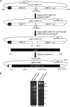
Engineering a full-length JHMV infectious BAC. (A) Strategy used to construct the JHMV BAC. First, a synthetic DNA was designed containing a CMV promoter, 1,991 bp of 5′ JHM sequence, and 1,197 bp of 3′ JHM sequence separated by a NotI site, and finally a poly(A) tail (pA), ribozyme cleavage site, HDV ribozyme, and BGH termination signal at the end. This DNA was then digested with SmaI and HindIII and ligated into the parental BAC plasmid, and the resulting clone was termed pBAC-JHMV 5′3′. Subsequently, pBAC-JHMV 5′3′ and a full-length JHMV ligated in vitro were digested with PacI and SanDI, ligated, and transformed into competent DH10B E. coli cells. The resulting BAC was termed pBAC-JHMV. (B) Confirmation of BAC clones. Representative clones of pBAC-JHMVIA and pBAC-JHMVSD were digested with EcoRI and EcoRV. The expected banding pattern for EcoRI digestion is 14.5, 11, 7.7, and 6.3 kb, while the expected pattern from EcoRV digestion is 22.3, 12.6, 4.7, and 0.2 kb. The resulting digests matched the expected digestion patterns (0.2-kb fragment is undetectable on the gel).
FIG 2
FIG 2
rJ virus engineered using pBAC-JHMV is infectious and replicates in a similar manner to other recombinant JHMVs. (A) Strategy for creating and recovering rJ virus from BAC clones. pBAC-JHMV DNA was transfected into DBT cells along with a plasmid expressing N protein. After plaques were visible (48 to 72 hpi), the virus was plaque purified two times and passaged two to three times on 17Cl-1 cells as described in Materials and Methods. (B and C) Recovered rJIA (B) and rJSD (C) viruses were analyzed for in vitro replication on 17Cl-1 cells. Progeny virus was collected at the indicated times postinfection, and yields were determined by plaque assay. p0, passage zero. Numbers in the legends indicate two distinct virus plaque isolates.
FIG 3
FIG 3
BAC-derived rJ retains the in vivo neurovirulence of other recombinant JHMV. Wild-type B6 mice were infected with 4E4 PFU rJIA (A and C) and 8E3 PFU rJSD (B and D) intranasally and monitored for survival and weight loss daily for 12 days. rJIA and rJSD controls refer to viruses constructed using targeted recombination and in vitro ligation, respectively. Data were combined from two independent experiments. rJSD 1.1, n = 5; rJIA control, rJSD control, and rJSD 1.2, n = 6; rJIA 5.1 and rJIA 5.3, n = 7. dpi, days postinfection.
FIG 4
FIG 4
rJ virus mutated at residue 1347 (N1347A) is minimally attenuated in vitro. (A) To compare replication kinetics of WT (GFP) and WT (GFP+) viruses, 17Cl-1 cells were infected at an MOI of 0.1 PFU/cell. Progeny virus was collected at the indicated times postinfection, and titers were determined by plaque assay. (B) pBAC-JHMV encoding the N1347A mutant was created using a two-step linear recombination with a Kanr-I-SceI dual marker cassette as described in Materials and Methods. Individually shaded boxes indicate regions of homology to viral sequence; the asterisk identifies the location of the active-site asparagine. Revertant rJ was also engineered (revN1347) using the same procedure. (C) Growth kinetics of rJIA. 17Cl-1 cells were infected with the indicated viruses. Progeny virus was collected at the indicated times postinfection, and yields were determined by plaque assay. (D) To assess RNA accumulation, 17Cl-1 cells were infected with the indicated viruses, and total RNA was collected at the indicated times postinfection. RNA was quantified by RT-qPCR with primers specific for genomic RNA and normalized to HPRT (Table 2 lists qPCR primers). The normalized amount of viral transcript at 6 hpi during revN1347 infection was set to 1. (E) Viral genome RNA/PFU ratio was determined by dividing the relative gRNA of revN1347 and N1347A virus from cells at 0 hpi by the PFU count of the viral stocks. The normalized ratio of revN1347 virus was set to 1. Data shown are mean values and standard errors of the means from two independent experiments with two different virus stocks performed in duplicate. (F) To examine viral fitness in vitro, 17Cl-1 cells were coinfected with N1347A (GFP+) and wild-type (GFP) virus or wild-type (GFP+) and wild-type (GFP) virus at a ratio of ∼4:1 and at a combined MOI of 0.1 PFU/ml. Virus was passaged three times in 17Cl-1 cells, with titers determined after each passage so that at each subsequent passage cells were infected at an MOI of 0.1 PFU/cell. The percentage of GFP-expressing virus was determined by dividing the number of GFP+ plaques by the number of total plaques. Passage 0 represents input virus. Data shown are mean values ± standard errors of the means (SEM) from a representative experiment performed in duplicate. *, P ≤ 0.05; **, P ≤ 0.005.
FIG 5
FIG 5
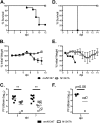
N1347A is attenuated in wild-type mice. (A and B) Wild-type B6 mice were infected intranasally with the indicated viruses and monitored daily for survival and weight loss. Weight curves were terminated at day 9 as most revN1347-infected mice had succumbed to infection by this point. Data are combined from two independent experiments (n = 13 in the revN1347 group and n = 14 in the N1347A group). (C) To determine virus load in the brains of infected mice, B6 mice were infected intranasally, and brains were homogenized at 5 and 7 days p.i. Homogenates were clarified by centrifugation, and titers were determined by plaque assay. Viral titers are expressed as the number of PFU/brain. Data were combined from two independent experiments (n = 7 per group at 5 days p.i. and n = 9 or 8 in the revN1347 or N1347A group, respectively, at 7 days p.i.). Lines represent mean values. (D and E) B6 mice were infected intracranially and monitored for survival and weight loss for 14 days (n = 3 mice per group). (F) Analysis of virus loads in the brains of intracranially infected mice. Brains were harvested at the indicated times and analyzed for viral loads as described for panel C. In panels C and F, the x axis indicates the limit of detection. Data were combined from two independent experiments (n = 4 per group). *, P ≤ 0.05; **, P ≤ 0.005.
FIG 6
FIG 6
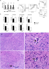
N1347A induces lower levels of proinflammatory cytokine and chemokine gene expression than revN1347 and reduced recruitment of immune cells into the brain and causes limited pathology in brains of infected mice. (A) To analyze proinflammatory cytokine and chemokine expression following infection, B6 mice were infected intranasally with revN1347 and N1347A viruses, and total brain RNA was collected at 5 days p.i. RNA was quantified by RT-qPCR with primers specific for CCL2, IFN-β, IL-6, TNF, and HPRT (Table 2 lists qPCR primers). The normalized amount of viral transcript in revN1347-infected mice was set to 1 (n = 6 for revN1347- and N1347A-infected mice; n = 3 for naive mice). Combined results from two independent experiments are shown. (B) Gating strategy for identifying leukocytes (CD45+), microglia (expressing intermediate levels of CD45 [CD45int] and CD11b+), macrophages (expressing high levels of CD45 [CD45high], CD11b+, and F4-80+), and neutrophils (CD45high CD11b+ Ly6C+ Ly6G+) from intranasally infected mice is shown. (C) Frequencies (upper panel) and cell numbers (lower panel) of total leukocytes, macrophages, microglia, and neutrophils are shown. Percentages of CD45+ cells are determined from the total cell population, while percentages for the individual cell populations refer to the CD45+ population (n = 5 for each group). Results are representative of two independent experiments. *, P ≤ 0.05; **, P ≤ 0.005. (D to G) Histological analysis of brains from mice infected intranasally with N1347A (D and E) or revN1347 (F and G) at 7 days p.i. Sections were prepared from the brainstems of infected mice and stained with hematoxylin and eosin. Foci of increased cellularity with apoptotic cells and cellular debris were detected primarily in revN1347-infected mice although perivascular infiltrates were found in mice infected with either virus. A region of the midbrain is shown in the figure. Black arrows indicate perivascular infiltrates. Original magnifications, ×100 (D and F) and ×400 (E and G). n = 3 for each group.
FIG 7
FIG 7
N1347A is attenuated in IFNAR−/− and TNF−/− mice. Eight-week-old IFNAR−/− (A and C) or 4- to 5-month-old TNF−/− (B and D) male mice were infected intranasally with revN1347 and N1347A viruses and monitored daily for survival and weight loss for 10 or 14 days, respectively, as indicated. For the experiments shown in panels A and C, n = 3 for the revN1347 infection group, and n = 5 for the N1347A infection group; for the experiments shown in panels B and D, n = 5 for the revN1347 infection group, and n = 4 for the N1347A infection group. Combined results from two independent experiments are shown. (E) N1347A does not have increased sensitivity to IFN compared to revN1347 virus. 17Cl-1 cells were pretreated with the indicated amounts of IFN-β (PBL, Piscataway, NJ) for 18 h, infected with revN1347 virus and N1347A virus at an MOI of 0.1 PFU/cell, and posttreated with IFN. Cells were analyzed for infectious virus at 20 hpi by plaque assay. Data are representative of two independent experiments.
FIG 8
FIG 8
N1347A is only modestly attenuated in RAG1−/− mice. (A and B) Two- to five-month-old RAG1−/− male mice were infected intranasally with revN1347 and N1347A and monitored daily for survival and weight loss. Combined results from three independent experiments are shown (n = 9 per group). (C) Brains from RAG1−/− mice infected with N1347A that were moribund or recovering were harvested at days 12 and 14, respectively, and viral titers were determined by plaque assay (n = 2 mice per group). The dashed line indicates the limit of detection.

References

    1. Perlman S, Netland J. 2009. Coronaviruses post-SARS: update on replication and pathogenesis. Nat Rev Microbiol 7:439–450. doi:10.1038/nrmicro2147. - DOI - PMC - PubMed
    1. Weiss SR, Navas-Martin S. 2005. Coronavirus pathogenesis and the emerging pathogen severe acute respiratory syndrome coronavirus. Microbiol Mol Biol Rev 69:635–664. doi:10.1128/MMBR.69.4.635-664.2005. - DOI - PMC - PubMed
    1. Neuman BW, Joseph JS, Saikatendu KS, Serrano P, Chatterjee A, Johnson MA, Liao L, Klaus JP, Yates JR III, Wuthrich K, Stevens RC, Buchmeier MJ, Kuhn P. 2008. Proteomics analysis unravels the functional repertoire of coronavirus nonstructural protein 3. J Virol 82:5279–5294. doi:10.1128/JVI.02631-07. - DOI - PMC - PubMed
    1. Han W, Li X, Fu X. 2011. The macro domain protein family: structure, functions, and their potential therapeutic implications. Mutation Res 727:86–103. doi:10.1016/j.mrrev.2011.03.001. - DOI - PMC - PubMed
    1. Feijs KL, Forst AH, Verheugd P, Luscher B. 2013. Macrodomain-containing proteins: regulating new intracellular functions of mono(ADP-ribosyl)ation. Nat Rev Mol Cell Biol 14:443–451. doi:10.1038/nrm3601. - DOI - PMC - PubMed

Publication types

MeSH terms