. 2015 Feb;89(3):1537-49.
doi: 10.1128/JVI.03306-14. Epub 2014 Nov 26.

Influenza A virus uses intercellular connections to spread to neighboring cells

Affiliations

Influenza A virus uses intercellular connections to spread to neighboring cells

Kari L Roberts et al. J Virol. 2015 Feb.

Abstract

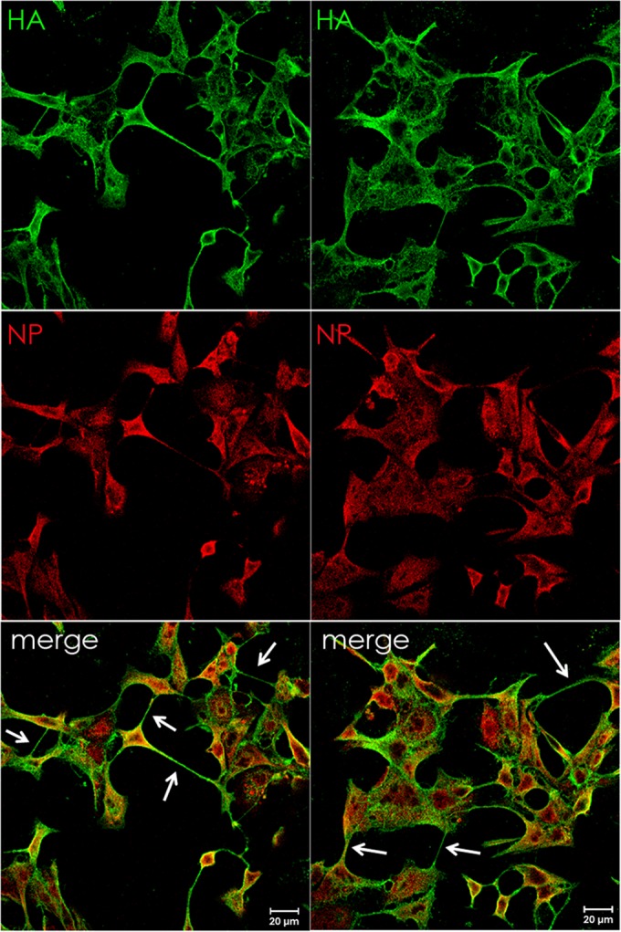
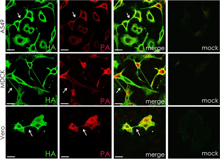
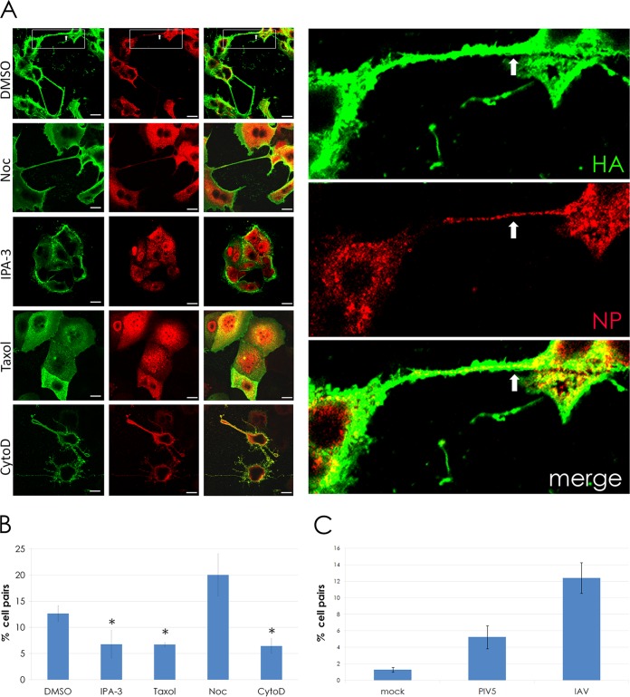
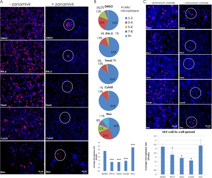
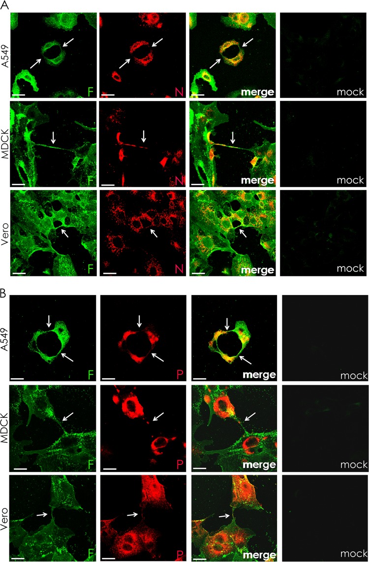
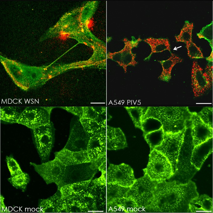
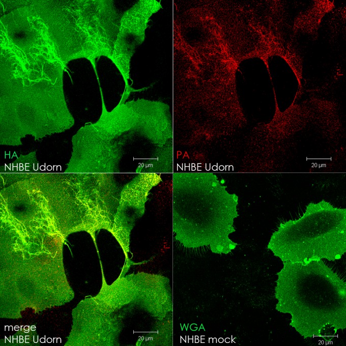
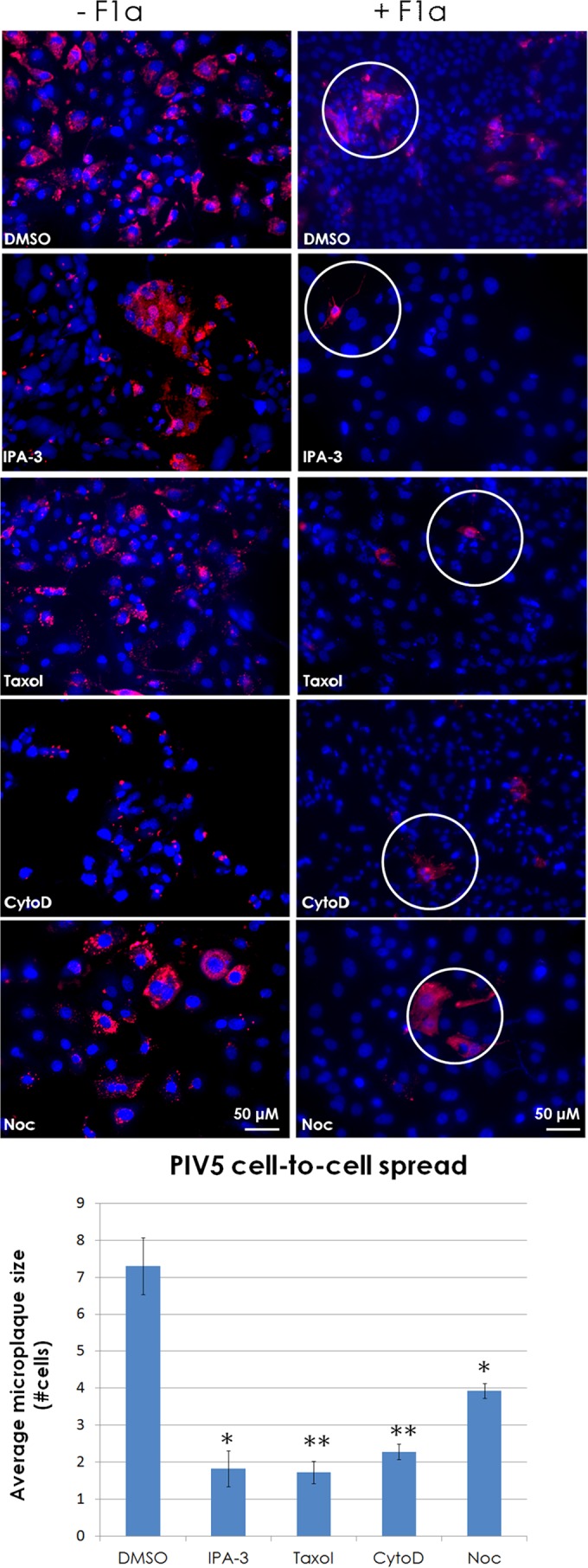
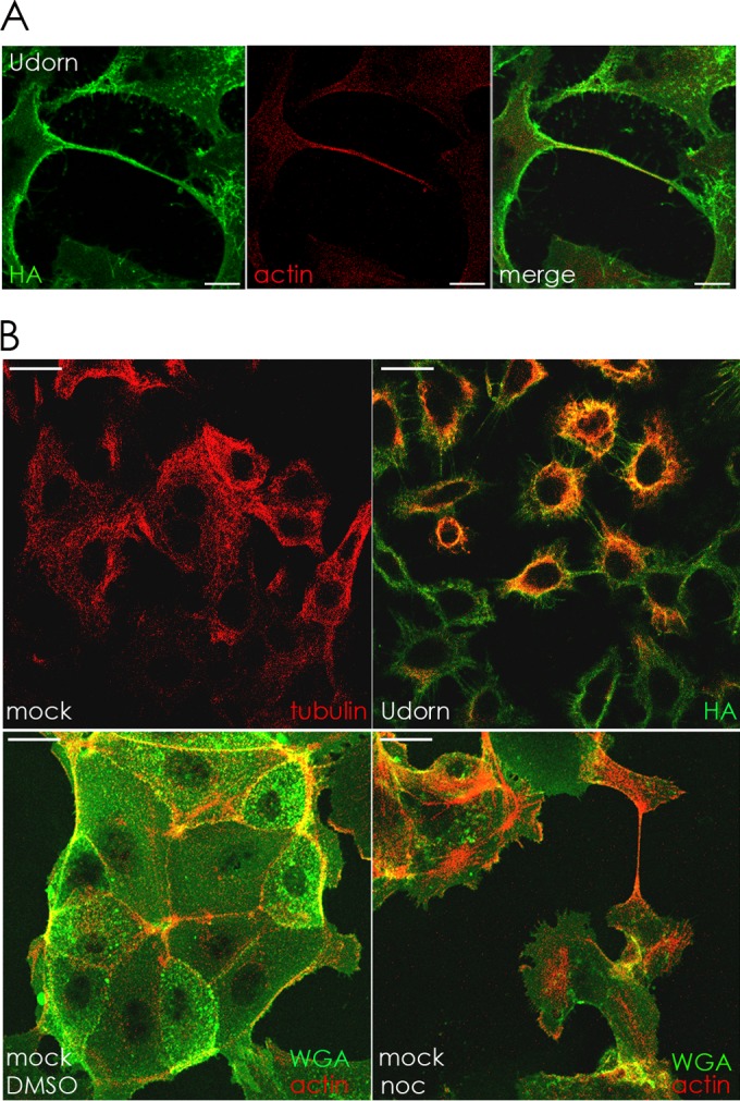
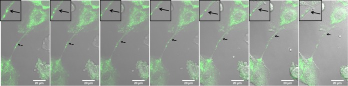
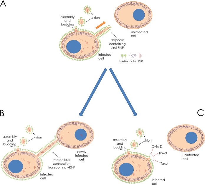
In the extracellular environment, cell-free virions seek out naive host cells over long distances and between organisms. This is the primary mechanism of spread for most viruses. Here we provide evidence for an alternative pathway previously undescribed for orthomyxoviruses, whereby the spread of influenza A virus (IAV) infectious cores to neighboring cells can occur within intercellular connections. The formation of these connections requires actin dynamics and is enhanced by viral infection. Connected cells have contiguous membranes, and the core infectious viral machinery (RNP and polymerase) was present inside the intercellular connections. A live-cell movie of green fluorescent protein (GFP)-tagged NS1 of IAV shows viral protein moving from one cell to another through an intercellular connection. The movement of tagged protein was saltatory but overall traveled only in one direction. Infectious virus cores can move from one cell to another without budding and release of cell-free virions, as evidenced by the finding that whereas a neuraminidase inhibitor alone did not inhibit the development of IAV microplaques, the presence of a neuraminidase inhibitor together with drugs inhibiting actin dynamics or the microtubule stabilizer paclitaxel (originally named taxol) precluded microplaque formation. Similar results were also observed with parainfluenza virus 5 (PIV5), a paramyxovirus, when neutralizing antibody was used to block spread by cell-free virions. Intercellular spread of infectious core particles was unaffected or enhanced in the presence of nocodazole for IAV but inhibited for PIV5. The intercellular connections have a core of filamentous actin, which hints toward transport of virus particles through the use of a myosin motor.

Importance: Here we describe a new method by which influenza A virus (IAV) spreads from cell to cell: IAV uses intracellular connections. The formation of these connections requires actin dynamics and is enhanced by viral infection and the absence of microtubules. Connected cells appeared to have contiguous membranes, and the core infectious viral machinery (RNP and polymerase) was present inside the intercellular connections. Infectious virus cores can move from one cell to another without budding and release of cell-free virions. Similar results were also observed with parainfluenza virus 5 (PIV5).


Figures

FIG 1
FIG 1
Viral ribonucleoprotein-containing intercellular connections form in IAV-infected cells. MDCK cells were infected with IAV (A/Udorn/72) at an MOI of 3 and fixed at 17 h p.i. Cell surfaces were immunostained for HA (green) and then permeabilized and immunostained for NP (red). Infected cells formed intercellular connections between neighboring cells that appeared to connect the cytoplasm of both cells. Images were photographed on a confocal microscope (Zeiss LSM 5).
FIG 2
FIG 2
Intercellular connections contain the IAV HA protein and a component (PA) of the polymerase in multiple cell types. A549, MDCK, and Vero cells were infected with IAV at an MOI of 3 and fixed at 17 h p.i. Cell surfaces were immunostained for hemagglutinin protein (HA; green) and then permeabilized and immunostained for viral polymerase (PA; red). Infected cells formed intracellular connections between neighboring cells that appeared to connect the cytoplasm of both cells. Images were photographed on a confocal microscope. Scale bar, 20 μm.
FIG 3
FIG 3
Intercellular connections formed during IAV infection require actin dynamics and F-actin but do not require microtubules and occur more frequently during infection. (A) MDCK cells were infected with IAV at an MOI of 3. At 1 h p.i., 30 μM IPA-3, 100 μM paclitaxel (“Taxol”), 20 μM cytochalasin D (CytoD), and 30 μM nocodazole (Noc) were added to infected cells, and the cells were fixed at 18 h p.i. Cell surfaces were immunostained for HA (green) and then permeabilized and immunostained for NP (red). Inset shows a zoomed image of two cells connected by an intercellular connection. Viral NP is clearly visible within the connection (arrows) as well as in the cell body. (B) The bar graph quantifies the percentage of MDCK cell pairs with intercellular connections in drug-treated and control (DMSO) cells infected with IAV. *, P < 0.05. (C) The bar graph quantifies the percentage of MDCK cell pairs connected by intercellular connections in mock, PIV5, or IAV infections. ***, P < 0.001. Images were photographed on a confocal microscope. Scale bar, 20 μm.
FIG 4
FIG 4
Intercellular connections provide a route for infection of neighboring cells. (A) MDCK cells were infected at an MOI of 0.1 with IAV. At 2 h p.i., 10 mM zanamivir, 30 μM IPA-3, 100 μM paclitaxel (“Taxol”), 20 μM cytochalasin D (CytoD), and 30 μM nocodazole (Noc) were added at 2 h p.i. as indicated, and the cells were incubated for 48 h. Cells were fixed and immunostained for NP (red), and nuclei were stained with DAPI (blue). Images were taken on a fluorescence microscope (Zeiss Axiovert 200M) using a 20× objective. (B) Quantification of the microplaques (NP-positive neighboring cells, white circles) in panel A. ***, P < 0.001. (C) MDCK cells were infected at an MOI of 0.1 with IAV and treated with or without 4 mM ammonium chloride and treated as described for panel A. Cells were fixed and immunostained as described for panel A. Microplaques (NP-positive neighboring cells, white circles) are quantified in a bar graph below the images. *, P < 0.05. Counts for 70 fields of view were included per sample per trial.
FIG 5
FIG 5
Intercellular connections form in paramyxovirus-infected cells and contain the RNP components N and P. A549, MDCK, and Vero cells were infected with PIV5 at an MOI of 3 and fixed at 24 h p.i. Cell surfaces were immunostained for the fusion protein (F; green) and then permeabilized and immunostained for either nucleoprotein (N, red) (A) or P protein (red) (B). Infected cells formed intercellular connections between neighboring cells that appeared to connect the cytoplasm of both cells. Images were photographed on a confocal microscope. Scale bar, 20 μm.
FIG 6
FIG 6
Intercellular connections form in multiple cell types infected with IAV (WSN) or PIV5. (Upper row) MDCK or A549 cells were infected with IAV A/WSN/33 or PIV5, respectively, at an MOI of 3. Cells were fixed at 17 h p.i. and immunostained for surface HA (WSN; green) or HN (PIV5; green) and then immunostained internally for NP (WSN; red) or P (PIV5; red). (Lower row) MDCK and A549 cells were mock infected and fixed 17 h later. Cell surfaces were stained with wheat germ agglutinin conjugated to Alexa Fluor 488 (5 μg/ml for 10 min). The white arrow indicates an example of vRNP-containing cell-cell connections. Images were photographed on a confocal microscope. Scale bar, 20 μm.
FIG 7
FIG 7
Intercellular connections form in IAV-infected primary cells. Normal human bronchial epithelial cells (NHBE) were infected with IAV (MOI = 1) or mock infected and fixed at 17 h p.i. in 10% formalin. IAV-infected cell surfaces were immunostained for hemagglutinin (HA; green), and then the cells were permeabilized and immunostained for polymerase (PA; red). Mock-infected cells were stained with Alexa Fluor 488-conjugated wheat germ agglutinin (WGA) to visualize the cell surface. Images were photographed on a confocal microscope. Scale bar, 20 μm.
FIG 8
FIG 8
Intercellular connections are utilized by the paramyxovirus parainfluenza virus 5 as a means of cell-to-cell spread. (A) MDCK cells were infected at an MOI of 0.1 with PIV5. At 2 h p.i., cells were treated with neutralizing MAb F1a or left untreated. Cells were then treated with or without IPA-3 (30 μM), paclitaxel (“Taxol”; 100 μM), cytochalasin D (CytoD; 20 μM), or nocodazole (Noc; 30 μM) and incubated for 48 h. Cells were fixed and immunostained for N protein (red), and nuclei were stained with DAPI (blue). Images were taken on a fluorescence microscope with a 20× objective. Microplaque (N-positive neighboring cells, white circles) quantification is shown in the bar graph. *, P < 0.05; **, P < 0.01. Counts for 70 fields of view were included per sample per trial.
FIG 9
FIG 9
(A) Intercellular connections contain filamentous actin. MDCK cells were infected with IAV at an MOI of 1 and fixed at 12 h p.i. in 10% formalin. Surface HA (green) was immunostained, and then the cells were permeabilized in 0.1% Tween–PBS and stained with phalloidin conjugated to Alexa Fluor 594 to visualize F-actin (red). Images were photographed on a confocal microscope. Scale bar, 20 μm. (B) IAV infection of cells affects microtubule formation, and depolymerization of microtubules in mock-infected cells does not inhibit formation of intercellular connections. Upper row, MDCK cells were mock infected or infected with IAV (MOI = 3) and fixed at 17 h p.i. Cell surfaces were immunostained with antibody to HA (green) and then permeabilized and immunostained with antibody to tubulin (red). Lower row, mock-infected MDCK cells were treated with DMSO or nocodazole (noc) and fixed 17 h later. Cells were stained with Alexa Fluor 488-conjugated WGA (green) to visualize cell surfaces and then permeabilized and stained with Alexa Fluor 594-conjugated phalloidin to visualize F-actin (red). Images were photographed on a confocal microscope. Scale bar, 20 μm.
FIG 10
FIG 10
Live-cell images show that GFP-tagged NS1 can be transported from one cell to another through an intercellular connection. MDCK cells were infected with IAV (MOI = 1) that expresses GFP-tagged NS1. Frames from a live-cell movie (see Movie S1 in the supplemental material) show GFP-tagged viral protein (NS1, arrows) moving through an intercellular connection and into the cytoplasm of a neighboring cell. Insets show a zoomed image of the area of interest for enhanced visualization of GFP-tagged protein and membrane border. Images were photographed on a confocal microscope. Scale bar, 20 μm.
FIG 11
FIG 11
Model for IAV spread via intercellular connections. (A) An IAV-infected cell neighboring an uninfected cell in the beginning stages of establishing an intercellular connection. Filamentous actin (purple) extends through the interior of the membrane-bound extension. (B) The cellular extension connects and fuses to the plasma membrane of the uninfected cell, allowing import of vRNP and establishment of infection. (C) Drugs inhibiting actin dynamics, F-actin, and a microtubule stabilizer (cytochalasin D [cyto D], IPA-3, and paclitaxel [Taxol], respectively) inhibit the formation of intercellular connections and therefore inhibit this form of cell-to-cell spread.

References

    1. Shaw ML, Palese P. 2013. Orthomyxoviridae, p 1151–1185 Fields virology, 6th ed Lippincott Williams & Wilkins, Philadelphia, PA.
    1. Stauffer S, Feng Y, Nebioglu F, Heilig R, Picotti P, Helenius A. 2014. Stepwise priming by acidic pH and a high K+ concentration is required for efficient uncoating of influenza A virus cores after penetration. J Virol 88:13029–13046. doi:10.1128/JVI.01430-14. - DOI - PMC - PubMed
    1. Amorim MJ, Bruce EA, Read EKC, Foeglein Á Mahen R, Stuart AD, Digard P. 2011. A Rab11- and microtubule-dependent mechanism for cytoplasmic transport of influenza A virus viral RNA. J Virol 85:4143–4156. doi:10.1128/JVI.02606-10. - DOI - PMC - PubMed
    1. Fontana J, Cardone G, Heymann JB, Winkler DC, Steven AC. 2012. Structural changes in influenza virus at low pH characterized by cryo-electron tomography. J Virol 86:2919–2929. doi:10.1128/JVI.06698-11. - DOI - PMC - PubMed
    1. Ye Z, Liu T, Offringa DP, McInnis J, Levandowski RA. 1999. Association of influenza virus matrix protein with ribonucleoproteins. J Virol 73:7467–7473. - PMC - PubMed

Publication types