. 2015 Feb;89(3):1879-88.
doi: 10.1128/JVI.03079-14. Epub 2014 Nov 26.

Nonmuscle myosin heavy chain IIb mediates herpes simplex virus 1 entry

Affiliations

Nonmuscle myosin heavy chain IIb mediates herpes simplex virus 1 entry

Jun Arii et al. J Virol. 2015 Feb.

Abstract

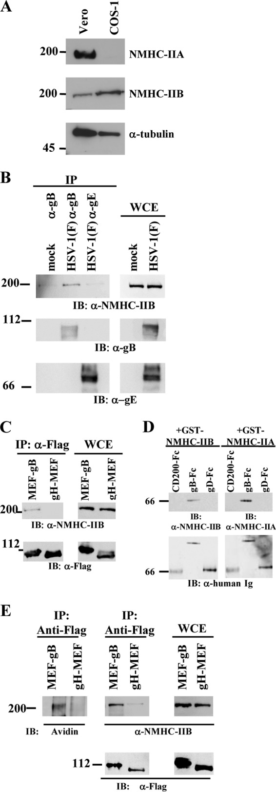
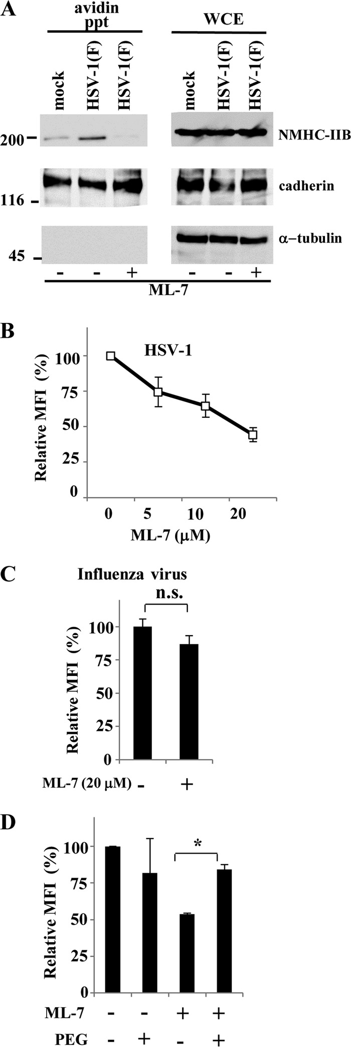
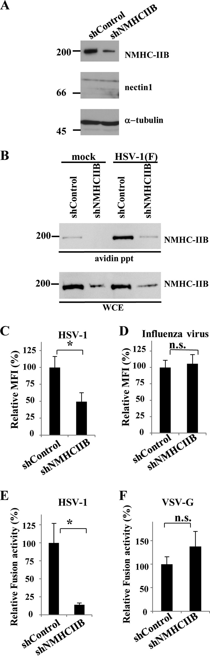
Nonmuscle myosin heavy chain IIA (NMHC-IIA) has been reported to function as a herpes simplex virus 1 (HSV-1) entry coreceptor by interacting with viral envelope glycoprotein B (gB). Vertebrates have three genetically distinct isoforms of the NMHC-II, designated NMHC-IIA, NMHC-IIB, and NMHC-IIC. COS cells, which are readily infected by HSV-1, do not express NMHC-IIA but do express NMHC-IIB. This observation prompted us to investigate whether NMHC-IIB might associate with HSV-1 gB and be involved in an HSV-1 entry like NMHC-IIA. In these studies, we show that (i) NMHC-IIB coprecipitated with gB in COS-1 cells upon HSV-1 entry; (ii) a specific inhibitor of myosin light chain kinase inhibited cell surface expression of NMHC-IIB in COS-1 cells upon HSV-1 entry as well as HSV-1 infection, as reported with NMHC-IIA; (iii) overexpression of mouse NMHC-IIB in IC21 cells significantly increased their susceptibility to HSV-1 infection; and (iv) knockdown of NMHC-IIB in COS-1 cells inhibited HSV-1 infection as well as cell-cell fusion mediated by HSV-1 envelope glycoproteins. These results supported the hypothesis that, like NMHC-IIA, NMHC-IIB associated with HSV-1 gB and mediated HSV-1 entry.

Importance: Herpes simplex virus 1 (HSV-1) was reported to utilize nonmuscle myosin heavy chain IIA (NMHC-IIA) as an entry coreceptor associating with gB. Vertebrates have three genetically distinct isoforms of NMHC-II. In these isoforms, NMHC-IIB is of special interest since it highly expresses in neuronal tissue, one of the most important cellular targets of HSV-1 in vivo. In this study, we demonstrated that the ability to mediate HSV-1 entry appeared to be conserved in NMHC-II isoforms. These results may provide an insight into the mechanism by which HSV-1 infects a wide variety of cell types in vivo.


Figures

FIG 1
FIG 1
Association of NMHC-IIB with HSV-1 gB. (A) Expression of NMHC-IIA and NMHC-IIB in Vero and COS-1 cells was analyzed by immunoblotting with anti-NMHC-IIA and anti-NMHC-IIB antibody, respectively. (B) COS-1 cells were mock incubated or incubated with wild-type HSV-1(F) at an MOI of 50 at 4°C for 2 h, followed by a temperature shift to 37°C for 2 min, and then immunoprecipitated (IP) with anti-gB or anti-gE antibody and analyzed by immunoblotting (IB) with anti-NMHC-IIB, anti-gB, or anti-gE antibody. WCE, whole-cell extract control. (C) COS-1 cells were incubated with YK711 (MEF-gB) or YK716 (gH-MEF) at an MOI of 50 at 4°C for 2 h, followed by a shift to 37°C for 2 min, and then immunoprecipitated with anti-Flag antibody and analyzed by immunoblotting with anti-NMHC-IIB antibody. WCE, whole-cell extract. (D) Purified GST-NMHC-IIA or GST-NMHC-IIB was reacted with purified gB-Fc, gD-Fc, or control Fc immobilized on protein A-Sepharose beads. The beads were then washed extensively and analyzed by immunoblotting with an anti-NMHC-IIA or -NMHC-IIB antibody. (E) Cell surface proteins of COS-1 cells exposed to YK711 (MEF-gB) or YK716 (gH-MEF) at 4°C for 2 h, followed by a temperature shift to 37°C for 15 min, were biotinylated, immunoprecipitated with an anti-Flag antibody, and analyzed by immunoblotting with streptavidin or anti-Flag or anti-NMHC-IIB antibody. Numbers at left of panels are molecular masses in kilodaltons.
FIG 2
FIG 2
Effect of ML-7 on cell surface expression of NMHC-IIB and HSV-1 infection in COS-1 cells. (A) Cell surface proteins of COS-1 cells incubated with wild-type HSV-1 at an MOI of 50 at 4°C for 2 h, followed by a temperature shift to 37°C for 15 min, in the presence or absence of 20 μM ML-7 were biotinylated, precipitated with avidin beads, and analyzed by immunoblotting with anti-NMHC-IIB or anticadherin or anti-α-tubulin antibody. Numbers at left are molecular masses in kilodaltons. (B and C) COS-1 cells infected with HSV-1 YK333 (EGFP) or influenza virus at an MOI of 1 in the presence of the indicated concentrations of ML-7 (B) or in the presence of 20 μM ML-7 (C) were analyzed by flow cytometry at 5 h and 7 h postinfection, respectively, and mean fluorescence intensities (MFIs) were determined. The mean and standard error of each data set are shown (n = 3; n.s., not significant). The mean value in the absence of ML-7 was normalized to 100% relative MFI. (D) COS-1 cells were infected with YK333 (EGFP) at an MOI of 1 in the absence or presence of 20 μM ML-7 for 1 h and subsequently treated with 40% PEG. Mean fluorescence intensities (MFIs) were determined by flow cytometry at 5 h postinfection. The relative MFI is the MFI of infected cells treated with ML-7 and/or PEG compared with untreated control cells. The mean and standard error of each data set are shown (n = 3; *, P = 0.0077, two-tailed Student t test). The mean value in the absence of ML-7 and PEG was normalized to 100% relative MFI.
FIG 3
FIG 3
Effect of NMHC-IIB overexpression on HSV-1 infection. (A) Expression of NMHC-IIB, NMHC-IIA, nectin-1, and α-tubulin in IC21/puro, IC21/hNMHC-IIA, IC21/mNMHC-IIB, and IC21/hnectin-1 cells was analyzed by immunoblotting. (B) Cell surface proteins of IC21/puro or IC21/mNMHC-IIB cells incubated with HSV-1 at an MOI of 50 at 4°C for 2 h, followed by a temperature shift to 37°C for 15 min, were biotinylated, precipitated with avidin beads, and analyzed by immunoblotting with anti-NMHC-IIB antibody. WCE, whole-cell extract. Numbers at left of panels A and B are molecular masses in kilodaltons. (C) IC21/puro, IC21/hNMHC-IIA, IC21/mNMHC-IIB, or IC21/hnectin-1 cells were infected with YK333 (EGFP) and analyzed by flow cytometry, and the percentage of infected cells was determined at 10 h postinfection. The mean and standard error of each data set are shown (n = 3; *, P < 0.01, two-tailed Student t test). (D) IC21/puro, IC21/hNMHC-IIA, IC21/mNMHC-IIB, or IC21/hnectin-1 cells were infected with YK333 (EGFP) at an MOI of 1 and analyzed by fluorescence and phase-contrast microscopy at 10 h postinfection.
FIG 4
FIG 4
Effects of NMHC-IIB knockdown on HSV-1 infection and cell-cell fusion. (A) NMHC-IIB, nectin-1, and α-tubulin expression levels in COS-shControl and COS-shNMHC-IIB cells were determined by immunoblotting. (B) Cell surface proteins of COS-shControl or COS-shNMHC-IIB cells incubated with HSV-1 at an MOI of 50 at 4°C for 2 h, followed by a temperature shift to 37°C for 15 min, were biotinylated, precipitated with avidin beads, and analyzed by immunoblotting with anti-NMHC-IIB antibody. WCE, whole-cell extract. Numbers at left of panels A and B are molecular masses in kilodaltons. (C and D) COS-shControl and COS-shNMHC-IIB cells infected with YK333 (EGFP) (C) or influenza virus (D) at an MOI of 1 were analyzed by flow cytometry, and mean fluorescence intensities (MFIs) were determined at 5 and 7 h postinfection, respectively. The mean and standard error of each data set are shown (n = 3; *, P = 0.036, n.s., not significant, two-tailed Student t test). The mean value for COS-shControl cells was normalized to 100% relative mean fluorescence intensity (MFI). (E and F) COS-1 cells transfected with expression vectors for T7 polymerase, gB, gD, gH, and gL (E) or expression vectors for T7 polymerase and vesicular stomatitis virus G protein (VSV-G) (F) were cocultured with COS-shControl or COS-shNMHC-IIB cells transfected with a reporter plasmid carrying the luciferase gene driven by the T7 polymerase. The mean and standard error of each data set are shown (n = 3; *, P = 0.034, two-tailed Student t test). The mean value for COS-shControl cells was normalized to 100% relative fusion activity.

References

    1. Roizman B, Knipe DM, Whitley RJ. 2007. Herpes simplex viruses, p 2501–2601 InKnipe DM, Howley PM, Griffin DE, Lamb RA, Martin MA, Roizman B, Strauss SE (ed), Fields virology, 5th ed Lippincott Williams & Wilkins, Philadelphia, PA.
    1. Herold BC, Visalli RJ, Susmarski N, Brandt CR, Spear PG. 1994. Glycoprotein C-independent binding of herpes simplex virus to cells requires cell surface heparan sulphate and glycoprotein B. J Gen Virol 75:1211–1222. doi:10.1099/0022-1317-75-6-1211. - DOI - PubMed
    1. Herold BC, WuDunn D, Soltys N, Spear PG. 1991. Glycoprotein C of herpes simplex virus type 1 plays a principal role in the adsorption of virus to cells and in infectivity. J Virol 65:1090–1098. - PMC - PubMed
    1. Spear PG. 2004. Herpes simplex virus: receptors and ligands for cell entry. Cell Microbiol 6:401–410. doi:10.1111/j.1462-5822.2004.00389.x. - DOI - PubMed
    1. Turner A, Bruun B, Minson T, Browne H. 1998. Glycoproteins gB, gD, and gHgL of herpes simplex virus type 1 are necessary and sufficient to mediate membrane fusion in a Cos cell transfection system. J Virol 72:873–875. - PMC - PubMed

Publication types

Substances