. 2014 Nov 26;34(48):15877-87.
doi: 10.1523/JNEUROSCI.2304-14.2014.

Proton-mediated block of Ca2+ channels during multivesicular release regulates short-term plasticity at an auditory hair cell synapse

Affiliations

Proton-mediated block of Ca2+ channels during multivesicular release regulates short-term plasticity at an auditory hair cell synapse

Soyoun Cho et al. J Neurosci. .

Abstract

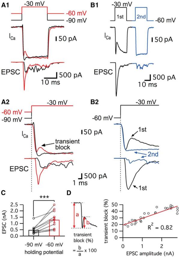
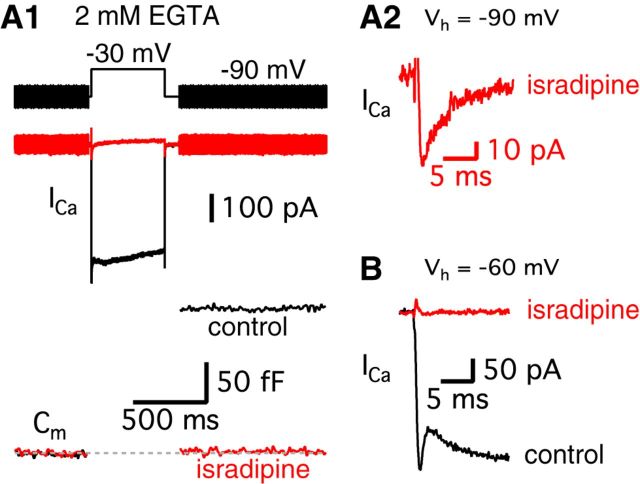
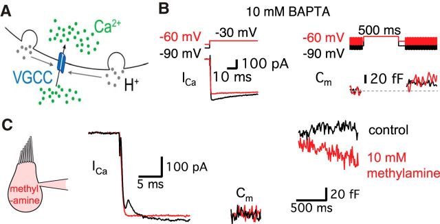
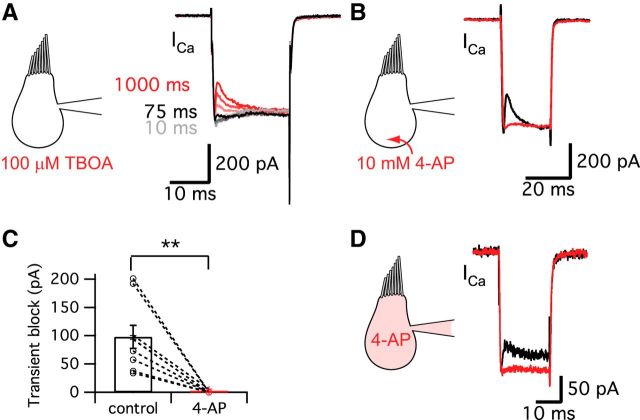
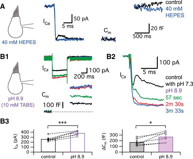
Synaptic vesicles release both neurotransmitter and protons during exocytosis, which may result in a transient acidification of the synaptic cleft that can block Ca(2+) channels located close to the sites of exocytosis. Evidence for this effect has been reported for retinal ribbon-type synapses, but not for hair cell ribbon synapses. Here, we report evidence for proton release from bullfrog auditory hair cells when they are held at more physiological, in vivo-like holding potentials (Vh = -60 mV) that facilitate multivesicular release. During paired recordings of hair cells and afferent fibers, L-type voltage-gated Ca(2+) currents showed a transient block, which was highly correlated with the EPSC amplitude (or the amount of glutamate release). This effect was masked at Vh = -90 mV due to the presence of a T-type Ca(2+) current and blocked by strong pH buffering with HEPES or TABS. Increasing vesicular pH with internal methylamine in hair cells also abolished the transient block. High concentrations of intracellular Ca(2+) buffer (10 mm BAPTA) greatly reduced exocytosis and abolished the transient block of the Ca(2+) current. We estimate that this transient block is due to the rapid multivesicular release of ∼600-1300 H(+) ions per synaptic ribbon. Finally, during a train of depolarizing pulses, paired pulse plasticity was significantly changed by using 40 mm HEPES in addition to bicarbonate buffer. We propose that this transient block of Ca(2+) current leads to more efficient exocytosis per Ca(2+) ion influx and it may contribute to spike adaptation at the auditory nerve.

Keywords: auditory; calcium current; electrophysiology; exocytosis; hair cells; protons.


Figures

Figure 1.
Figure 1.
Ca2+ currents of bullfrog hair cells exhibit a transient block at a holding potential of −60 mV. A, Paired whole-cell voltage-clamp recordings of presynaptic hair cell and a connected afferent fiber. The hair cell was depolarized from −90 mV (black) or −60 mV (red) to −30 mV for 20 ms (A1). When a hair cell was held at −60 mV, the postsynaptic afferent fiber showed a larger EPSC than for hair cells at −90 mV (A2). At −60 mV, the Ca2+ current showed a transient block in contrast with Ca2+ currents of hair cells at −90 mV (A2). EPSCs evoked by stimulating hair cells from −60 mV (red) showed shorter synaptic delay than that for hair cells at −90 mV (black) (A2). B, When the hair cells were stimulated by a pair of 20 ms pulses from −60 to −30 mV, the transient block was observed in the first Ca2+ current (black in B1) with large EPSCs and was absent in the second Ca2+ current (blue in B1). With strong paired-pulse depression, the second Ca2+ current does not show the transient block. B2 shows overlapped traces of Ca2+ currents with EPSCs from B1 on an expanded time scale. C, The peak amplitudes of EPSCs were significantly increased by changing the holding potential of hair cells from −90 mV (485 ± 104 pA) to −60 mV (1292 ± 208 pA) within the same pairs of recordings (n = 11, p < 0.001, paired t test). Open circles indicate individual pairs. D, The ratio of transient block in Ca2+ currents was measured from the amplitude of transient block (b) divided by the peak amplitude of Ca2+ currents (a). The ratio of transient block strongly correlates with the amplitude of the EPSCs (n = 27 measurements from 7 pairs). The red line shows a linear fit (slope = 0.010 ± 0.001, R2 = 0.82).
Figure 2.
Figure 2.
The transient block of the Ca2+ current requires a certain period of time at the more depolarized holding potential of −60 mV. A, Ca2+ currents were recorded from hair cells held at −90 mV using a 20-ms-long pulse to −30 mV followed by pre-depolarization to −60 mV for 10–1000 ms. Different colors represent different durations of the pre-depolarization from −90 to −60 mV. Ca2+ currents started showing a transient block with 75 ms of pre-depolarization (arrow). B, A depolarization from −90 to −60 mV evokes a small, non-inactivating Ca2+ current in the hair cell. C, Ca2+ currents elicited by the 20 ms pulses with various pre-depolarization (10, 75, 200, and 1000 ms) were superimposed. The transient block becomes clearly larger as the duration of the pre-depolarization increases.
Figure 3.
Figure 3.
T-type Ca2+ currents mask the transient block for hair cells held at −90 mV. A1, A hair cell was depolarized from −90 to −30 mV for 500 ms in control (black) and after perfusion of 10 μm isradipine, a specific L-type Ca2+channel blocker (red). The standard intracellular hair cell Ca2+ buffer was 2 mm EGTA. 10 μm isradipine strongly inhibited Ca2+ currents and the Cm jump elicited by the depolarizing pulse. A2, After applying isradipine, a small Ca2+ current still persists in hair cells held at −90 mV (red). B, After holding hair cells at −60 mV, isradipine completely blocked the Ca2+ current (red).
Figure 4.
Figure 4.
The transient block of Ca2+ current requires the exocytosis of synaptic vesicles. A, Diagram showing that H+ (gray) ions are released from fused vesicles during exocytosis and can affect adjacent Ca2+ channels at an active zone of release (blue). B, After the dialysis of 10 mm BAPTA into a hair cell, Ca2+ currents were evoked by a 500 ms pulse from −90 mV (black) or from −60 mV (red) to −30 mV. Note that Ca2+ currents do not show the transient block now that exocytosis has been severely decreased by 10 mm BAPTA. C, 10 mm methylamine was added to the internal pipette solution. This removed the transient block, which went from 81 ± 15 to 0 pA (n = 6, p = 0.0028, paired t test) without significant changes in ΔCm (136 ± 2.4 fF in control and 126 ± 4.8 fF in 10 mm methylamine, n = 6, p > 0.05, paired t test). The Ca2+ current was evoked by a 500-ms-long pulse from a holding potential of −60 to −30 mV. The Ca2+ currents were recorded in control (black) with a first patch pipette and then, after repatching the same hair cell, using a second pipette containing 10 mm methylamine (red).
Figure 5.
Figure 5.
Glutamate transporter and K+ currents do not contribute to the transient block. A, Ca2+ currents were evoked in hair cells held at −90 mV using a pre-depolarization to −60 mV for 10–1000 ms and then by a 20-ms-long pulse to −30 mV (same protocol as Fig. 2A). The Ca2+ currents for the 20 ms pulses are shown superimposed. The transient block of the Ca2+ currents was not blocked by 100 μm TBOA, a specific blocker of glutamate transporters (n = 6). The different colors represent different durations of the pre-depolarization. The Ca2+ current started showing the transient block within ∼75 ms of the pre-depolarization, which was very similar to control (Fig. 2C). B, 4-AP removed the transient block of the Ca2+ current. However, this effect may not be related with the blocking of K+ currents, but rather is probably due to the H+ capturing ability of 4-AP and its possible disruption of normal vesicle pH. C, 10 mm 4-AP significantly decreased the amplitude of transient block from 98 ± 20 pA to 0.6 ± 0.6 pA (n = 9). **p < 0.01, paired t test. Open circles indicate individual pairs. D, When hair cells were depolarized from −60 to −30 mV for 20 ms, 10 mm 4-AP in the patch pipette solution significantly decreased the transient block from 52.0 ± 9.4 pA (black) to 2.2 ± 2.1 pA (red, n = 9, p = 0.0011, paired t test). Control recordings were performed within 27 ± 7 s (n = 9) after break-in and the recordings of 4-AP effects were done at 4 min 39 ± 30 s (n = 9) after break-in to whole-cell mode.
Figure 6.
Figure 6.
Protons released during exocytosis cause a transient block of Ca2+ currents. A, The hair cell was depolarized from −60 to −30 mV for 500 ms in control condition (black) and after perfusion of external solution including 40 mm HEPES (blue). B, External solution of pH 8.9 with 10 mm TABS increased the amplitude of Ca2+ currents and the ΔCm jump, but inhibited the transient block of Ca2+ currents. Initial kinetics of the Ca2+ currents in B1 are shown in an expanded time scale in B2. As a pH 8.9 solution was applied, the transient block of Ca2+ currents was quickly removed. The time lapsed after a control recording as the pH 8.9 solution was being perfused is shown in different colors. In B3, we quantify the effects of the external solution with pH 8.9. The amplitudes of Ca2+ currents (left) and ΔCm (right) evoked by a 500-ms-long depolarization from −60 to −30 mV are compared in control (black) and after >3–5 min of perfusion with the pH 8.9 solution (purple). The average amplitude of Ca2+ currents was significantly increased from 262 ± 22 to 383 ± 20 pA (n = 6) and ΔCm was significantly enhanced from 174 ± 26 to 271 ± 39 fF (n = 6) by pH 8.9 external solution. *p < 0.05, paired t test; ***p = 0.001, paired t test. Open circles indicate individual pairs.
Figure 7.
Figure 7.
Effects of released H+ ions on EPSC amplitudes during a 400 Hz train of stimuli. A, Hair cells were stimulated by a train of 1 ms depolarizing pulses from −60 to −30 mV with an interpulse interval of 1.5 ms. A total of 40 stimuli were given at 400 Hz for 100 ms. Ca2+ currents showed a transient block with a double exponential recovery (dashed line; τfast = 3.9 ms and τslow = 23.7 ms) in control (left). EPSCs evoked by the 400 Hz train of pulses showed depression (dashed line; τfast = 1.3 ms and τslow = 7.4 ms). After perfusion of the external solution with added 40 mm HEPES (right), the hair cell Ca2+ currents do not show the transient block and the corresponding EPSCs show depression with a single exponential time constant (τ = 2.6 ms). B, EPSCs in control (black) and 40 mm HEPES (red) were normalized and superimposed. The second and third EPSCs become larger when 40 mm HEPES removed the transient block in the Ca2+ currents. Note that the second EPSC (arrow) was significantly larger with 40 mm HEPES than in control. C, The ratio of the first and the second EPSCs (EPSC2/EPSC1) in control (black) and with 40 mm HEPES (red). EPSC2/EPSC1 with 40 mm HEPES was significantly increased (0.42 ± 0.04) compared with that in control (0.28 ± 0.03; n = 6). D, From 6 pairs, the average τ of the EPSC peaks in 40 mm HEPES (red, 2.6 ± 0.3 ms) was significantly increased compared with control (black, 1.4 ± 0.2 ms). E, When hair cells were stimulated by a 400 Hz train of 1 ms depolarizing pulses from −60 to −30 mV, an external solution with 10 mm 4-AP removed the transient block in Ca2+ currents and slowed the kinetics of the EPSC train depression (fit with a single exponential time constant: τ = 2.5 ms). F, EPSC2/EPSC1 in control (black) and with 10 mm 4-AP (red). EPSC2/EPSC1 with 10 mm 4-AP was significantly increased (0.45 ± 0.06) compared with that in control (0.28 ± 0.05; n = 5). G, From 5 pairs, the average τ of the EPSC peaks in 10 mm 4-AP (red, 3.5 ± 0.6 ms) was significantly increased compared with control (black, 1.8 ± 0.2 ms). *p < 0.05, paired t test; **p < 0.01, paired t test. Open circles indicate individual pairs.

References

    1. Armstrong CM, Loboda A. A model for 4-Aminopyridine action on K channels: Similarities to tetraethylammonium ion action. Biophys J. 2001;81:895–904. doi: 10.1016/S0006-3495(01)75749-9. - DOI - PMC - PubMed
    1. Awatramani GB, Price GD, Trussell LO. Modulation of transmitter release by presynaptic resting potential and background calcium levels. Neuron. 2005;48:109–121. doi: 10.1016/j.neuron.2005.08.038. - DOI - PubMed
    1. Ballanyi K, Kaila K. Activity-evoked changes in intracellular pH. In: Kaila K, Ransom BR, editors. pH and Brain Function. New York: Wiley; 1998. pp. 291–308.
    1. Barnes S, Merchant V, Mahmud F. Modulation of transmission gain by protons at the photoreceptor output synapse. Proc Natl Acad Sci U S A. 1993;90:10081–10085. doi: 10.1073/pnas.90.21.10081. - DOI - PMC - PubMed
    1. Beg AA, Ernstrom GG, Nix P, Davis MW, Jorgensen EM. Protons act as a transmitter for muscle contraction in C. elegans. Cell. 2008;132:149–160. doi: 10.1016/j.cell.2007.10.058. - DOI - PMC - PubMed

Publication types

LinkOut - more resources