. 2014 Nov 26;34(48):16140-52.
doi: 10.1523/JNEUROSCI.1908-14.2014.

PMP22 is critical for actin-mediated cellular functions and for establishing lipid rafts

Affiliations

PMP22 is critical for actin-mediated cellular functions and for establishing lipid rafts

Sooyeon Lee et al. J Neurosci. .

Abstract

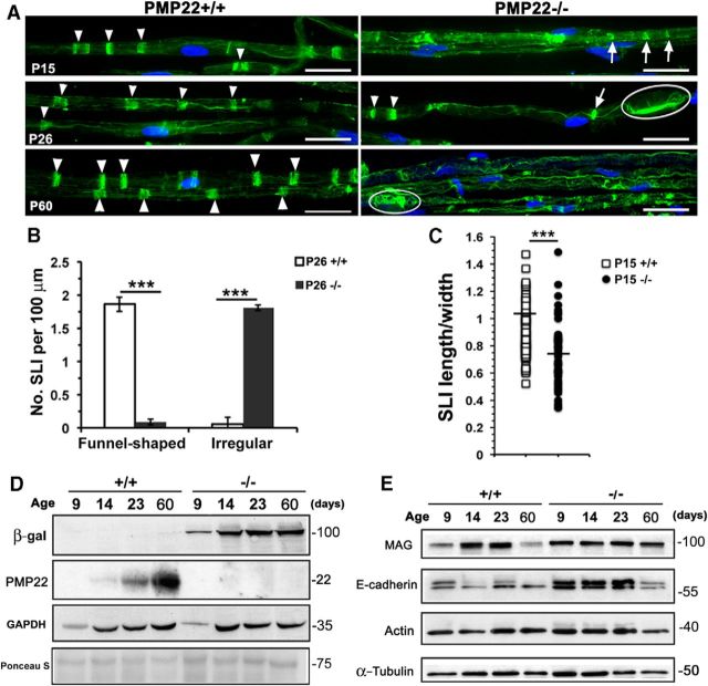
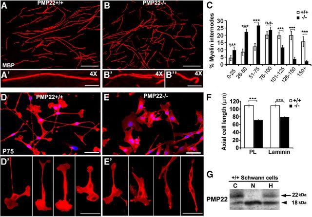
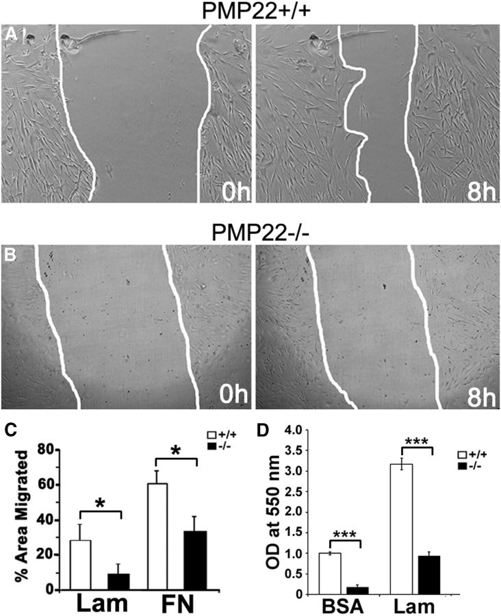
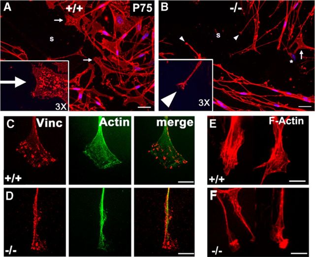
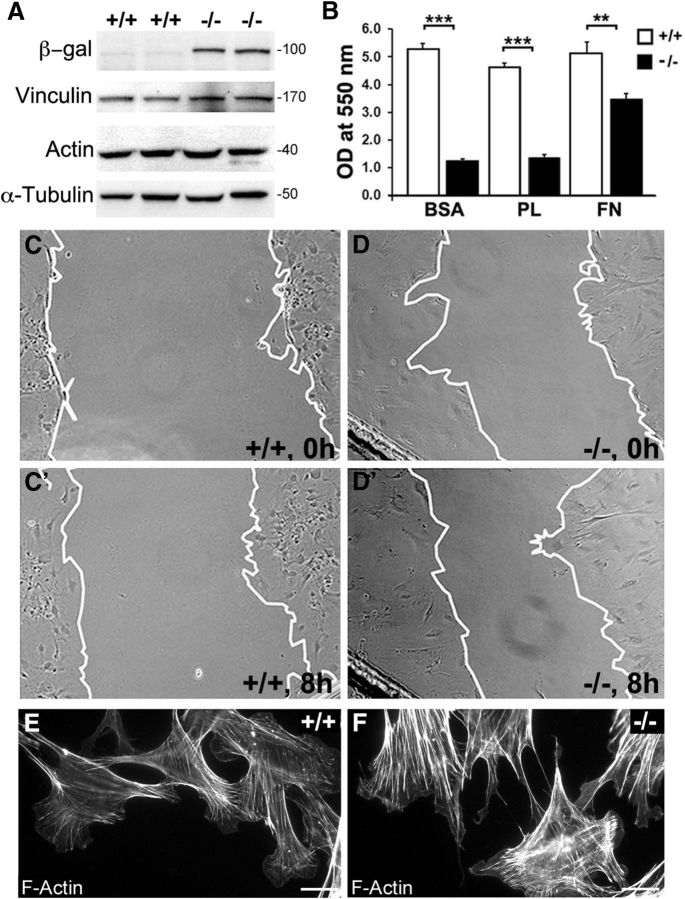
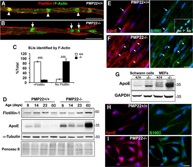
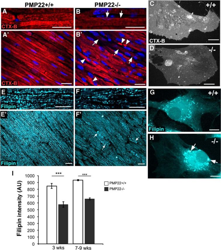
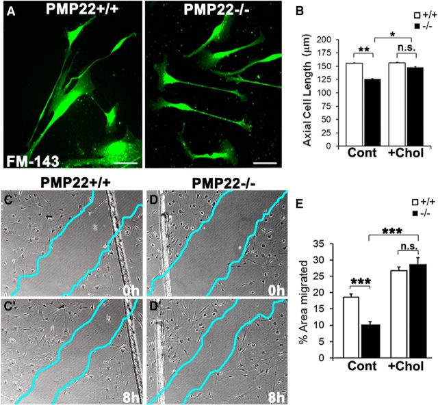
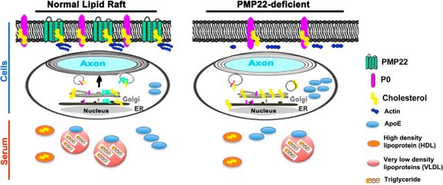
Haploinsufficiency of peripheral myelin protein 22 (PMP22) causes hereditary neuropathy with liability to pressure palsies, a peripheral nerve lesion induced by minimal trauma or compression. As PMP22 is localized to cholesterol-enriched membrane domains that are closely linked with the underlying actin network, we asked whether the myelin instability associated with PMP22 deficiency could be mediated by involvement of the protein in actin-dependent cellular functions and/or lipid raft integrity. In peripheral nerves and cells from mice with PMP22 deletion, we assessed the organization of filamentous actin (F-actin), and actin-dependent cellular functions. Using in vitro models, we discovered that, in the absence of PMP22, the migration and adhesion capacity of Schwann cells and fibroblasts are similarly impaired. Furthermore, PMP22-deficient Schwann cells produce shortened myelin internodes, and display compressed axial cell length and collapsed lamellipodia. During early postnatal development, F-actin-enriched Schmidt-Lanterman incisures do not form properly in nerves from PMP22(-/-) mice, and the expression and localization of molecules associated with uncompacted myelin domains and lipid rafts, including flotillin-1, cholesterol, and GM1 ganglioside, are altered. In addition, we identified changes in the levels and distribution of cholesterol and ApoE when PMP22 is absent. Significantly, cholesterol supplementation of the culture medium corrects the elongation and migration deficits of PMP22(-/-) Schwann cells, suggesting that the observed functional impairments are directly linked with cholesterol deficiency of the plasma membrane. Our findings support a novel role for PMP22 in the linkage of the actin cytoskeleton with the plasma membrane, likely through regulating the cholesterol content of lipid rafts.

Keywords: Schwann cell; actin cytoskeleton; cholesterol; lipid rafts; myelin; neuropathy.


Figures

Figure 1.
Figure 1.
Alterations in F-actin and proteins of Schmidt-Lanterman incisures in the absence of PMP22. A, Sciatic nerves from WT (PMP22+/+) and PMP22−/− mice at the indicated ages were labeled with phalloidin to identify F-actin. Funnel-shaped normal incisures (arrowheads), irregularly shaped incisures (arrows), and tomaculae (circle) are marked. Nuclei are labeled with Hoechst dye (blue). Scale bars, 25 μm. B, Quantification of funnel-shaped and irregular SLIs per unit length of nerve (n = 5–6 individual nerve samples per genotype). C, Scatter plot of SLI length/width index from P26 sciatic nerves (n = 5–6 individual nerve samples per genotype). D, E, Sciatic nerve total protein lysates (15 μg/lane) were immunoblotted with the indicated antibodies. Ages of the donor tissues are indicated in postnatal days. GAPDH, α-tubulin, and Ponceau S stains of the blots are shown as protein-loading controls. Molecular weight is reported in kilodaltons (right).
Figure 2.
Figure 2.
PMP22 augments the elongation of myelinating and nonmyelinating Schwann cells. A, B, Representative images of MBP-like immunoreactivity in myelinating DRG explants from WT (PMP22+/+) (A) and PMP22−/− (B) mice. A, B, B, 4× magnification of MBP-positive myelinated internodes. C, Frequency distribution of internodes lengths is graphed. Internode lengths were binned into 25 μm intervals and graphed as a percentage of the total number of internodes. D–E, Morphology of WT (PMP22+/+) and PMP22−/− Schwann cells after immunolabeling with anti-P75 antibody (red). Nuclei were stained with Hoechst dye and are shown in blue. D′–E, Representative individual WT and PMP22−/− Schwann cells are shown. F, Measurements of axial cell length after anti-P75 immunolabeling and plating on PL or laminin. G, Total protein lysates (30 μg/lane) of normal Schwann cells were digested with N-glycosidase (N) or endoH (H) and were probed with anti-PMP22 antibody. Lane C contains samples incubated without enzyme. Arrows mark the glycosylated PMP22, while arrowheads point to the deglycosylated PMP22. Molecular mass is reported in kilodaltons. Values represent the mean ± SEM. ***p < 0.001, two-tailed Student's t test. n.s, Not significant. For the quantifications, data were analyzed from three independent cultures. Scale bars: A, B, 100 μm; DE, 20 μm.
Figure 3.
Figure 3.
PMP22 regulates Schwann cell migration and adhesion. A, B, Representative phase contrast images of WT (PMP22+/+) and PMP22−/− Schwann cells at 0 and 8 h postscratch in the wound migration assay. White lines indicate wound boundaries. C, Quantification of area migrated by WT and PMP22−/− cells on laminin (Lam) and on fibronectin (FN). D, Adhesion assay of Schwann cells on BSA or Lam. Data are graphed after optical density (OD) values were normalized to the BSA controls and represent the mean ± SD. The data shown are representative of three to five independent culture experiments. Student's t test: *p < 0.05; ***p < 0.001.
Figure 4.
Figure 4.
Lack of PMP22 in Schwann cells is associated with morphological alterations in lamellipodial spreading. A, B, Schwann cells at the wound edge in WT (PMP22+/+) and PMP22−/− cultures after immunolabeling with anti-P75 antibody at 8 h postscratch. s, scratch area; arrows, lamellipodia; arrowheads, filopodia. Insets in A and B show a 3× enlarged view of representative lamellipodia. C, D, Images of lamellipodia of WT and PMP22−/− Schwann cells after colabeling with anti-vinculin and anti-actin antibodies. E, F, Micrographs of WT and PMP22−/− Schwann cell processes after labeling with phalloidin. Images are representative of cells from three to four independent cultures. Scale bars: A, B, 20 μm; C–F, 10 μm.
Figure 5.
Figure 5.
PMP22 is critical for adhesion and migration in fibroblasts. A, Total protein extracts (20 μg/lane) from WT (PMP22+/+) and PMP22−/− fibroblasts were blotted with antibodies against β-gal, vinculin, and actin. α-Tubulin is shown as a protein-loading control. Molecular mass is reported in kilodaltons. B, Adhesion assay with WT and PMP22−/− fibroblasts on BSA, PL, or fibronectin (FN)-coated wells. Values represent the mean ± SEM. Student's t test: **p < 0.01; ***p < 0.001. n = 3 independent culture experiments, with duplicate wells per experiment. C–D, Representative phase contrast images of WT and PMP22−/− fibroblasts at 0 h (C, D) and 8 h (C, D) after scratch wounding. The migration front is indicated with a white line. E, F, F-actin labeling with phalloidin after 8 h scratch wound migration in WT and PMP22−/− fibroblasts. At the migration front, PMP22−/− cells (F) display prominent stress fibers compared with WT controls (E). Scale bars: E, F, 20 μm.
Figure 6.
Figure 6.
The absence of PMP22 perturbs the localization of the raft protein flotillin-1 and is associated with an increase in Apo E expression. A, B, Sciatic nerves from P15 WT (PMP22+/+) and PMP22−/− mice were double labeled with phalloidin (green) and anti-flotillin-1 antibody (red). Arrowheads (A) indicate flotillin-1 immunoreactivity at F-actin-enriched SLIs, while arrows (B) point to SLIs with F-actin-lacking surrounding flotillin-1. Scale bars, 20 μm. C, Quantification of SLIs flanked by flotillin-1 in nerve samples from WT and PMP22−/− mice. ***p < 0.001, Student's t test. n = 5–6 nerves per genotype. D, Total sciatic nerve lysates (20 μg/lane) from PMP22+/+ and PMP22−/− mice at the indicated ages were immunoblotted with antibodies against flotillin-1 and ApoE. α-Tubulin and Ponceau S stains of the membrane are shown as controls for protein loading. Molecular weight is reported in kilodaltons (right). E, F, ApoE and S-100β coimmunostaining of WT and PMP22−/− sciatic nerves are shown. Arrows indicate ApoE-like immunoreactivity within Schwann cells, identified by S-100β. Nuclei are shown in blue, after staining with Hoechst dye. Inset in E is an image of a WT nerve section with the anti-mouse secondary antibody alone. G, Western blot of ApoE on lysates from WT and PMP22−/− Schwann cells and MEFs. GAPDH is shown as a protein-loading control. H, I, Double immunolabeling of cultured Schwann cells with ApoE and S-100β. Nuclei are shown in blue with Hoechst dye. Scale bar, 20 μm.
Figure 7.
Figure 7.
The absence of PMP22 perturbs lipid rafts. A–B, Alexa Fluor 594-conjugated Ctx-β (red) and Hoechst dye (blue) labeling on sciatic nerves from 3- to 4-week-old WT and PMP22−/− mice. A′–B, low-magnification images of the nerves shown in A and B. In samples from PMP22−/− mice, arrows (B, B) indicate reduced Ctx-β binding, while arrowheads (B) mark localized clusters of Ctx-β. C, D, Cell surface labeling of normal (C) and PMP22-deficient (D) cultured Schwann cells with Ctx-β. E–F, Visualization of cholesterol-filipin on sciatic nerve sections from 3- to 4-week-old WT and PMP22−/− mice. Lower-magnification views of the nerves are shown in E and F. G, H, Filipin staining of PMP22+/+ and PMP22−/− Schwann cells. Arrows indicate enlarged clusters of cholesterol in PMP22−/− Schwann cells. Scale bars: A–D, 20 μm; E–H, 10 μm. I, Quantification of filipin fluorescence intensity in nerves from 3- and 7- to 9-week-old WT and PMP22−/− mice is shown. AU, Arbitrary units. ***p < 0.001, Student's t test. n = 3 nerves per genotype.
Figure 8.
Figure 8.
The addition of cholesterol rescues the migration and morphology defects of PMP22-deficient Schwann cells. A, PMP22+/+ and PMP22−/− Schwann cells labeled with FM143 after the addition of 20 μg/ml cholesterol for 24 h. Scale bars, 10 μm. B, Axial cell length measured in normal and PMP22−/− Schwann cells in the presence of cholesterol (+Chol). C, D, Representative 10× phase contrast images of control (Cont) PMP22+/+ (C) and PMP22−/− Schwann cells (D) before and after 8 h of migration in the presence of 20 μg/ml cholesterol. The migration front is shown as blue lines. E, Quantification of the percentage of the area migrated in the absence or presence of exogenous cholesterol. B, E, Values represent the mean ± SEM; *p < 0.05; **p < 0.01; ***p < 0.001; n.s., not significant; two-tailed Student's t test. Data were obtained from two to three independent cell culture experiments.
Figure 9.
Figure 9.
Model of how the absence of PMP22 leads to alterations in the localization of lipid raft-associated filamentous actin and perturbation in cholesterol homeostasis. In the normal nerve, lipid rafts are enriched in cholesterol, and contain PMP22 and protein 0 (P0). Filamentous actin, comprising SLIs, are anchored at lipid rafts. Levels of cholesterol and the cholesterol-binding protein ApoE are low within myelinated Schwann cells. In the absence of PMP22, the nerves are depleted in cholesterol, while ApoE expression is elevated within Schwann cells. Vascular plasma cholesterol levels are stable, while serum ApoE levels are reduced. In the absence of PMP22, the lipid balance of the nerve–vascular unit is altered.

Similar articles

Cited by

References

    1. Abramoff MD, Magalhães PJ, Ram SJ. Image processing with ImageJ. Biophotonics Int. 2004;11:36.
    1. Adlkofer K, Martini R, Aguzzi A, Zielasek J, Toyka KV, Suter U. Hypermyelination and demyelinating peripheral neuropathy in Pmp22-deficient mice. Nat Genet. 1995;11:274–280. doi: 10.1038/ng1195-274. - DOI - PubMed
    1. Amici SA, Dunn WA, Jr, Murphy AJ, Adams NC, Gale NW, Valenzuela DM, Yancopoulos GD, Notterpek L. Peripheral myelin protein 22 is in complex with α6β4 integrin, and its absence alters the Schwann cell basal lamina. J Neurosci. 2006;26:1179–1189. doi: 10.1523/JNEUROSCI.2618-05.2006. - DOI - PMC - PubMed
    1. Amici SA, Dunn WA, Jr, Notterpek L. Developmental abnormalities in the nerves of peripheral myelin protein 22-deficient mice. J Neurosci Res. 2007;85:238–249. doi: 10.1002/jnr.21118. - DOI - PubMed
    1. Aumailley M, Mann K, von der Mark H, Timpl R. Cell attachment properties of collagen type VI and Arg-Gly-Asp dependent binding to its alpha 2(VI) and alpha 3(VI) chains. Exp Cell Res. 1989;181:463–474. doi: 10.1016/0014-4827(89)90103-1. - DOI - PubMed

Publication types