

Diagnostic challenges in primary orbital fibrosarcoma: a case report
- PMID: 25429202
- PMCID: PMC4242685
- DOI: 10.2147/OPTH.S70843
Diagnostic challenges in primary orbital fibrosarcoma: a case report
Abstract
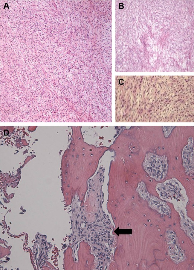
Fibrosarcoma is a rare and malignant spindle cell tumor of mesenchymal origin that infrequently presents in the orbit. Evolving diagnostic criteria confound the identification of these tumors, as well as the interpretation of previous studies on this unusual entity. We report a case of a 66-year-old man with a mass on his left inferior orbit, with associated paresthesia. A computed tomography (CT) scan showed a lesion on the left anteroinferomedial orbit, with bone erosion. An en bloc surgical excision followed by a thorough immunohistologic evaluation allowed diagnosis of an orbital fibrosarcoma. The patient has had no recurrence after 14 months of follow up. Once a commonly identified soft tissue malignancy, fibrosarcoma has become a diagnosis of exclusion as a result of improved diagnostic and classification techniques, such as immunohistochemistry and fluorescence in situ hybridization (FISH). This type of soft tissue tumor is now an uncommon entity, and we report the first case of a primary orbital fibrosarcoma in an adult, using modern diagnostic and classification methods.
Keywords: fibroblasts; mesenchymal tumor; soft tissue malignancy; spindle cell orbital tumor.
Figures


References
-
- Duke-Elder S, MacFaul PA, editors. System of Ophthalmology Vol XIII: The Ocular Adnexa Part II: Lacrimal, Orbital, and Para-orbital Diseases. London: Henry Kimpton; 1974.
-
- Rootman J. Diseases of the Orbit. A Multidisciplinary Approach. 2nd ed. Philadelphia, PA: Lippincott Williams and Wilkins; 2003.
-
- Bahrami A, Folpe AL. Adult-type fibrosarcoma: A reevaluation of 163 putative cases diagnosed at a single institution over a 48-year period. Am J Surg Pathol. 2010;34(10):1504–1513. - PubMed
-
- Coindre JM. Grading of soft tissue sarcomas: review and update. Arch Pathol Lab Med. 2006;130(10):1448–1453. - PubMed
-
- Folpe AL. Fibrosarcoma: a review and update. Histopathology. 2014;64(1):12–25. - PubMed
Publication types
LinkOut - more resources
Full Text Sources
Other Literature Sources
Research Materials
