. 2014 Nov 12:8:387.
doi: 10.3389/fnbeh.2014.00387. eCollection 2014.

Abnormal emotional learning in a rat model of autism exposed to valproic acid in utero

Affiliations

Abnormal emotional learning in a rat model of autism exposed to valproic acid in utero

Anwesha Banerjee et al. Front Behav Neurosci. .

Abstract

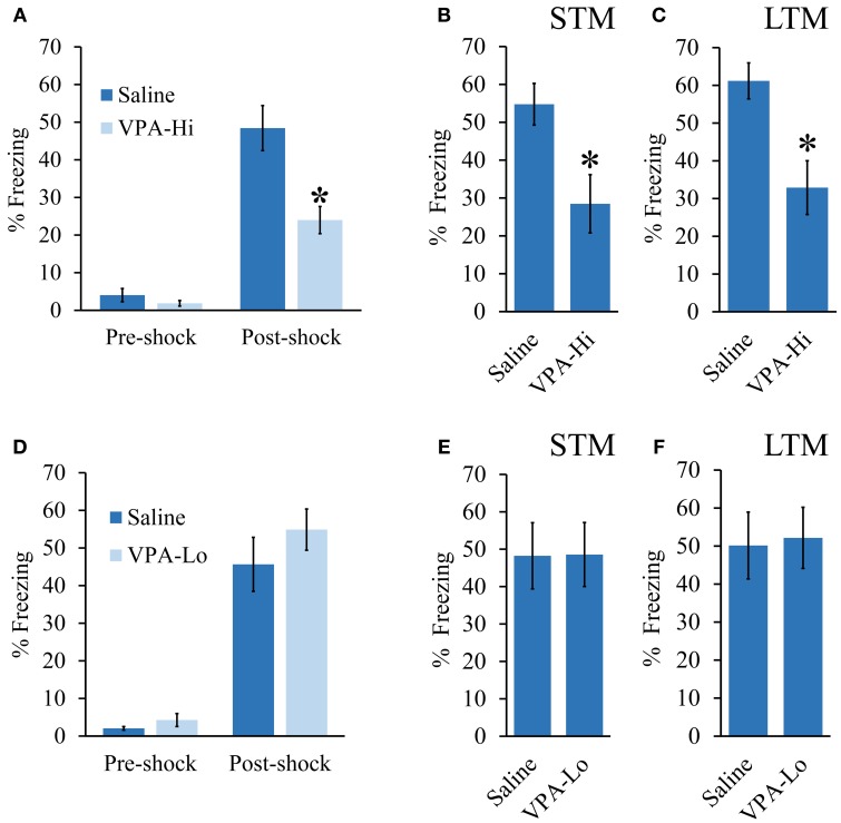
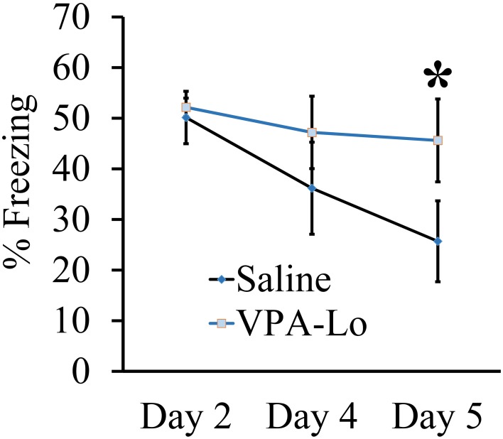
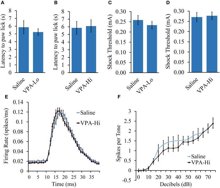
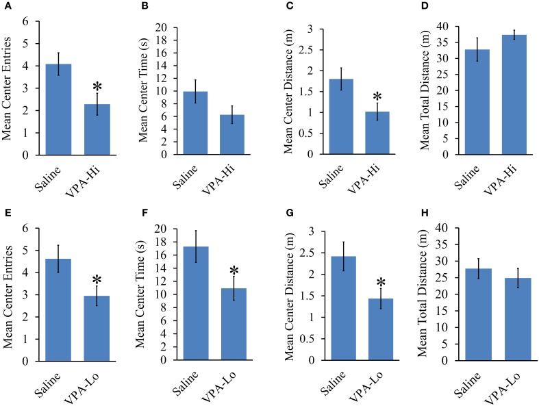
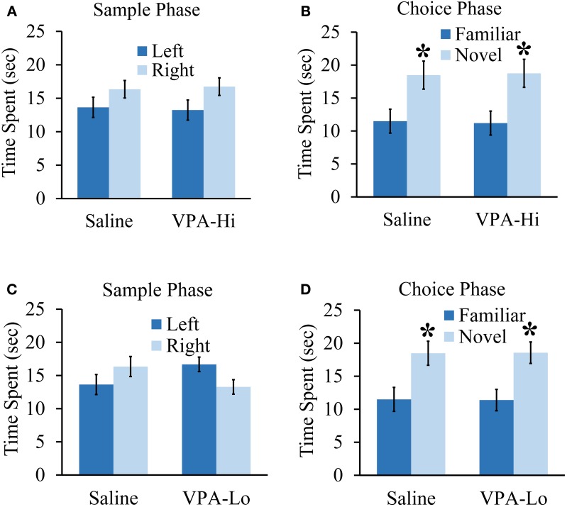
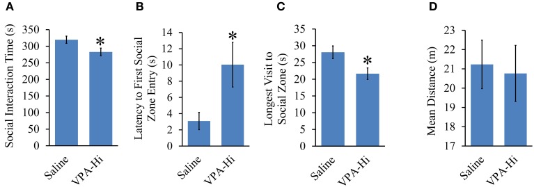
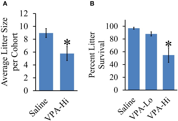
Autism Spectrum Disorders (ASD) are complex neurodevelopmental disorders characterized by repetitive behavior and impaired social communication and interactions. Apart from these core symptoms, a significant number of ASD individuals display higher levels of anxiety and some ASD individuals exhibit impaired emotional learning. We therefore sought to further examine anxiety and emotional learning in an environmentally induced animal model of ASD that utilizes the administration of the known teratogen, valproic acid (VPA) during gestation. Specifically we exposed dams to one of two different doses of VPA (500 and 600 mg/kg) or vehicle on day 12.5 of gestation and examined the resultant progeny. Our data indicate that animals exposed to VPA in utero exhibit enhanced anxiety in the open field test and normal object recognition memory compared to control animals. Animals exposed to 500 mg/kg of VPA displayed normal acquisition of auditory fear conditioning, and exhibited reduced extinction of fear memory and normal litter survival rates as compared to control animals. We observed that animals exposed to 600 mg/kg of VPA exhibited a significant reduction in the acquisition of fear conditioning, a significant reduction in social interaction and a significant reduction in litter survival rates as compared to control animals. VPA (600 mg/kg) exposed animals exhibited similar shock sensitivity and hearing as compared to control animals indicating the fear conditioning deficit observed in these animals was not likely due to sensory deficits, but rather due to deficits in learning or memory retrieval. In conclusion, considering that progeny from dams exposed to rather similar doses of VPA exhibit striking differences in emotional learning, the VPA model may serve as a useful tool to explore the molecular and cellular mechanisms that contribute to not only ASD, but also emotional learning.

Keywords: Pavlovian fear conditioning; amygdala; autism; emotion; learning; memory; valproic acid.


Figures

Figure 1
Figure 1
Progeny from VPA exposed dams exhibit a dose dependent effect on fear learning: (A–C) Progeny from VPA-Hi and saline exposed dams were auditory fear conditioned (Saline = 16, VPA-Hi = 13). (A) Pre and post shock freezing was assessed immediately before and after exposure to tone-shock, respectively. (B) Auditory fear memory assessed 3 h post fear conditioning (i.e., STM) (C) Auditory fear memory assessed 24 h post fear conditioning (i.e., LTM). (D–F) Progeny from VPA-Lo and saline exposed dams were auditory fear conditioned (Saline = 12, VPA-Lo = 12). (D) Pre and post shock freezing was assessed immediately before and after exposure to tone-shock, respectively. (E) Auditory fear memory assessed 3 h post fear conditioning (i.e., STM) (F) Auditory fear memory assessed 24 h post fear conditioning (i.e., LTM). Bars represent the mean ± standard error of the mean (SEM) (*p < 0.05).
Figure 2
Figure 2
Progeny from VPA-Lo exposed dams exhibit reduced fear extinction to a conditioned auditory cue. Progeny from VPA-Lo and saline exposed dams were auditory fear conditioned and LTM/extinction learning was assessed on days 2, 4, and 5 post fear conditioning (Saline = 12, VPA-Lo = 12). Bars represent the mean ± s.e.m. (*p < 0.05).
Figure 3
Figure 3
The fear conditioning deficit observed in progeny from VPA-Hi exposed dams is likely not due to deficits in sensory modalities necessary for auditory fear conditioning. (A) Thermal nociception as measured by the hotplate test did not differ between progeny from VPA-Lo and saline exposed dams (Saline = 12, VPA-Lo = 12) (B) or progeny from VPA-Hi and saline exposed dams (Saline = 12, VPA-Lo = 11) (C) Response to foot shock stimuli did not differ between progeny from VPA-Lo and saline exposed dams (Saline = 10, VPA-Lo = 10) (D) or progeny from VPA-Hi and saline exposed dams (Saline = 12, VPA-Lo = 11). (E) The primary auditory cortex response strength and response latency to a 5 kHz tone did not differ between progeny from VPA-Hi and saline exposed dams (Saline = 10, VPA-Hi = 10). (F) Hearing ability to a 5 kHz, 0–75dB tone did not differ between progeny from VPA-Hi and saline exposed dams (Saline = 10, VPA-Hi = 10) as measured by auditory cortex neuronal spike firing upon exposure to 5 kHz, 0–75 dB tones. Data presented as mean ± s.e.m.
Figure 4
Figure 4
Progeny from VPA exposed dams exhibit increased anxiety and normal locomotor behavior. Progeny from VPA-Hi dams exhibit increased anxiety in an open field as indicated by, (A) a reduced number of center entries, (B) a trend of reduced center time, (C) a reduced center distance traveled, without affecting, (D) the mean total distance traveled as compared to saline exposed animals (Saline = 25, VPA-Hi = 21). Progeny from VPA-Lo exposed dams exhibit increased anxiety in an open field as indicated by, (E) a reduced number of center entries, (F) a reduced center time, (G) a reduced center distance traveled, without affecting, (H) the mean total distance traveled as compared to saline exposed animals (Saline = 21, VPA-Lo = 20). Data presented as mean ± s.e.m. (*p < 0.05).
Figure 5
Figure 5
Progeny from VPA exposed dams exhibit normal objection recognition memory. (A) Progeny from VPA-Hi and saline exposed dams spent equal time with the familiar object during the sample phase of an object recognition test (saline = 10, VPA-Hi = 8). (B) VPA-Hi and saline animals spent more time than chance with a novel object during the choice phase of an object recognition test, indicating normal retention of object memory. (C) The VPA-Lo group had a slight preference for the object situated on left side compared to right side (saline = 10, VPA-Lo = 10) (saline = 10, VPA-Lo = 10). (D) VPA-Lo and saline exposed animals spent more time than considered chance with a novel object during the choice phase of an object recognition test, indicating normal retention of object memory. Data presented as mean ± s.e.m. time spent with each object (*p < 0.05).
Figure 6
Figure 6
Progeny from VPA-Hi exposed dams exhibit reduced social interaction. (A) VPA-Hi animals spent less time in the social interaction zone compared to saline animals (saline = 15, VPA-Hi = 15). (B) Latency to first entry into the social interaction zone was higher in VPA-Hi animals compared to saline animals. (C) The longest visit to the social interaction zone was reduced in VPA-Hi animals. (D) Mean distance traveled in the social interaction apparatus did not differ significantly between the groups. Data presented as mean ± s.e.m. (*p < 0.05).
Figure 7
Figure 7
Effect of VPA exposure in utero on average litter size and percent litter survival. (A) VPA-Hi animals exhibited a significant reduction in average litter size per cohort in comparison to saline animals. (B) The percent litter survival was reduced significantly in VPA-Hi animals compared to saline and VPA-Lo animals. No significant difference in percent litter survival was observed in VPA-Lo animals compared to saline animals. Data presented as mean ± s.e.m. (*p < 0.05).

References

    1. Adachi M., Autry A. E., Covington H. E., 3rd., Monteggia L. M. (2009). MeCP2-mediated transcription repression in the basolateral amygdala may underlie heightened anxiety in a mouse model of Rett syndrome. J. Neurosci. 29, 4218–4227. 10.1523/JNEUROSCI.4225-08.2009 - DOI - PMC - PubMed
    1. Amir R. E., Van Den Veyver I. B., Wan M., Tran C. Q., Francke U., Zoghbi H. Y. (1999). Rett syndrome is caused by mutations in X-linked MECP2, encoding methyl-CpG-binding protein 2. Nat. Genet. 23, 185–188. 10.1038/13810 - DOI - PubMed
    1. Arndt T. L., Stodgell C. J., Rodier P. M. (2005). The teratology of autism. Int. J. Dev. Neurosci. 23, 189–199. 10.1016/j.ijdevneu.2004.11.001 - DOI - PubMed
    1. Bachevalier J., Loveland K. A. (2006). The orbitofrontal-amygdala circuit and self-regulation of social-emotional behavior in autism. Neurosci. Biobehav. Rev. 30, 97–117. 10.1016/j.neubiorev.2005.07.002 - DOI - PubMed
    1. Bailey A., Le Couteur A., Gottesman I., Bolton P., Simonoff E., Yuzda E., et al. (1995). Autism as a strongly genetic disorder: evidence from a British twin study. Psychol. Med. 25, 63–77. 10.1017/S0033291700028099 - DOI - PubMed