Review
. 2014 Oct;2(5):10.1128/microbiolspec.MGM2-0006-2013.
doi: 10.1128/microbiolspec.MGM2-0006-2013.

Mycobacterium tuberculosis Serine/Threonine Protein Kinases

Affiliations
Review

Mycobacterium tuberculosis Serine/Threonine Protein Kinases

Sladjana Prisic et al. Microbiol Spectr. 2014 Oct.

Abstract

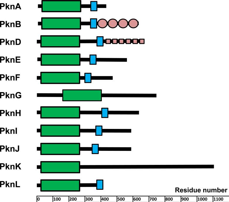
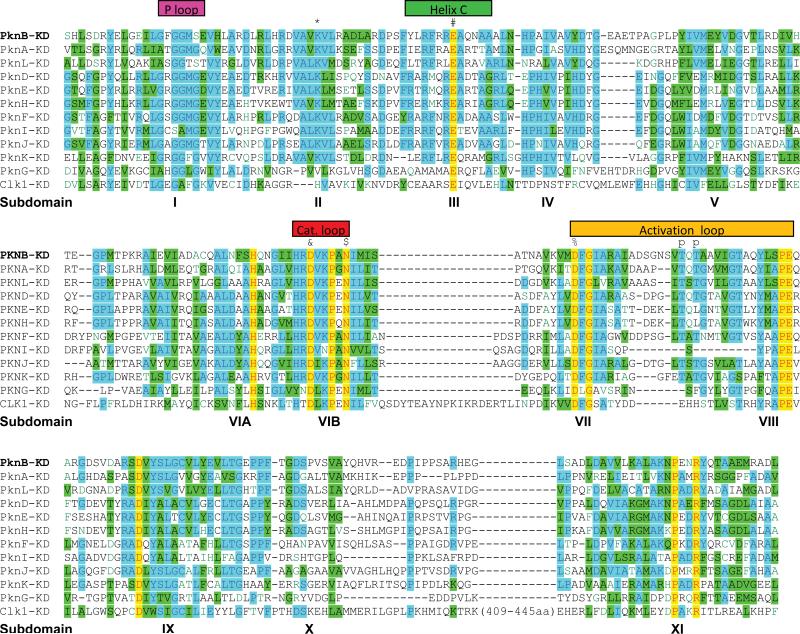
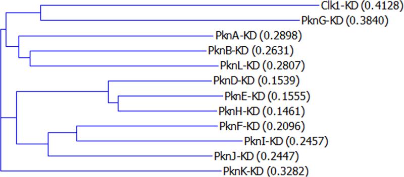
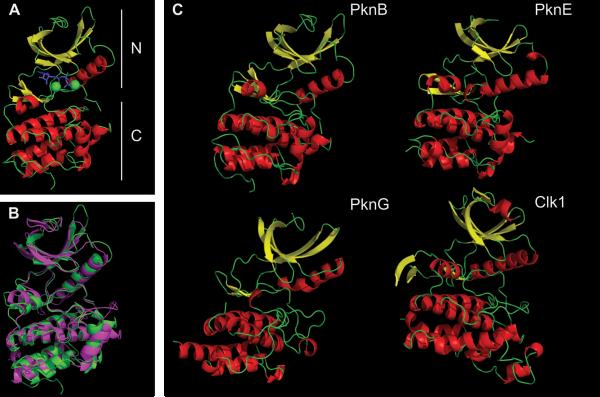
The Mycobacterium tuberculosis genome encodes 11 serine/threonine protein kinases (STPKs). A similar number of two-component systems are also present, indicating that these two signal transduction mechanisms are both important in the adaptation of this bacterial pathogen to its environment. The M. tuberculosis phosphoproteome includes hundreds of Ser- and Thr-phosphorylated proteins that participate in all aspects of M. tuberculosis biology, supporting a critical role for the STPKs in regulating M. tuberculosis physiology. Nine of the STPKs are receptor type kinases, with an extracytoplasmic sensor domain and an intracellular kinase domain, indicating that these kinases transduce external signals. Two other STPKs are cytoplasmic and have regulatory domains that sense changes within the cell. Structural analysis of some of the STPKs has led to advances in our understanding of the mechanisms by which these STPKs are activated and regulated. Functional analysis has provided insights into the effects of phosphorylation on the activity of several proteins, but for most phosphoproteins the role of phosphorylation in regulating function is unknown. Major future challenges include characterizing the functional effects of phosphorylation for this large number of phosphoproteins, identifying the cognate STPKs for these phosphoproteins, and determining the signals that the STPKs sense. Ultimately, combining these STPK-regulated processes into larger, integrated regulatory networks will provide deeper insight into M. tuberculosis adaptive mechanisms that contribute to tuberculosis pathogenesis. Finally, the STPKs offer attractive targets for inhibitor development that may lead to new therapies for drug-susceptible and drug-resistant tuberculosis.


Figures

FIGURE 1
FIGURE 1
Domain organization of STPKs from M. tuberculosis. Domains were predicted using the SMART algorithm (, ). Kinase domains are shown as green boxes, trans-membrane portions are in blue, and some known extracellular domains are in light red. doi:10.1128/microbiolspec.MGM2-0006-2013.f1
FIGURE 2
FIGURE 2
Sequence alignment of STPKs from M. tuberculosis. Kinase domains predicted by the SMART algorithm (see Fig. 1) were aligned and grouped using AlignX software (Life Technologies). Human Clk1 kinase is also included for comparison. Major features are noted. Selected conserved residues are labeled with the following symbols (residue numbers from PknB): *, Lys40; #, Glu59; &, Asp138; $, Asn143; %, Asp156; p, major phosphorylation sites in the activation loop. doi:10.1128/microbiolspec.MGM2-0006-2013.f2
FIGURE 3
FIGURE 3
Dendrogram of KDs of M. tuberculosis STPKs. KDs identified by the SMART algorithm were aligned and grouped using the AlignX software (Life Technologies). Human Clk1 kinase is also included for comparison. Distance scores as given by AlignX are shown in parentheses. doi:10.1128/microbiolspec.MGM2-0006-2013.f3
FIGURE 4
FIGURE 4
Overview of the M. tuberculosis STPK's KD. (A) Major features of the PknB KD (1MRU_B): N-terminal (upper) and C-terminal (lower) lobes are labeled. The ATP analog is in blue, and two Mg2+ ions are in green. (B) Overlap of PknB (green) and Clk1 (magenta). Clk1 was a top hit when the PknB structure was used to search similar three-dimensional structures using the NCBI VAST program. For clarity, residues 298 to 319 and 395 to 443 in Clk1 that are absent in M. tuberculosis STPKs (see Fig. 2) are truncated in Clk1. (C) PknB (1MRU_B), PknE (2H34_B), PknG (2PZI_A), and Clk1 (1Z57). α-Helix is in red, β-sheet is in yellow. Figures were made using PyMOL (Schrödinger) and POV-Ray (povray.org). doi:10.1128/microbiolspec.MGM2-0006-2013.f4
FIGURE 5
FIGURE 5
Active site of PknB KD. (A) Overlap of “closed” PknB KD (1MRU_B) in green and “open” apo-PknE-KD (2H34) in blue, with the PknE C helix labeled in red. (B) PknB active site (1MRU_B) P loop (GFGGMS), magenta; Mg2+, red balls; ATPγS, yellow; C-helix, green (Glu59-green); Lys40, aqua; catalytic loop, red (Asp138-orange, Asn143-red); DFG motif, purple (Asp156-purple). Figures were made using PyMOL (Schrödinger) and POV-Ray (povray.org). doi:10.1128/microbiolspec.MGM2-0006-2013.f5
FIGURE 6
FIGURE 6
Back-to-back dimerization of KDs. (A) PknB-KD dimer showing “back-to-back” interaction. (B) Overlap of PknB-KD in active form (1MRU_B-blue) and conformations of the PknB-KD L33D mutant that perturbs the dimer interface (3ORK, yellow; 3ORI_A, red; 3ORL, green). The C helix is shown in ribbon, while the rest of the structure is shown in wire. (C) C helix from the PknB structures in panel B magnified to highlight differences in the position of Glu59. Figures were made using PyMOL (Schrödinger) and POV-Ray (povray.org). doi:10.1128/microbiolspec.MGM2-0006-2013.f6
FIGURE 7
FIGURE 7
Distinct modes of monomer interaction in dimers of PknB versus PknG (A) “Front-to-front” dimer of mutant PknB KD (3F69) in complex with Kt5720 inhibitor (yellow). The “substrate” subunit (magenta) has most of its activation loop disordered (red), while the “enzyme” subunit (blue) has a well-defined activation loop (orange) with visible phosphor-ylated Thr171 (green). (B) Structure of PknG (2PZI) in complex with inhibitor Ax20017 (magenta). Three domains: rubredoxin (yellow), KD (green), and TPR domain (red) are shown only in one subunit. The second subunit is depicted in gray. Figures were made in PyMOL (Schrödinger) and POV-Ray (povray.org). doi:10.1128/microbiolspec.MGM2-0006-2013.f7
FIGURE 8
FIGURE 8
M. tuberculosis phosphoproteome. Phosphoproteins were identified in all functional categories of M. tuberculosis proteins (). doi:10.1128/microbiolspec.MGM2 -0006-2013.f8

References

    1. West AH, Stock AM. Histidine kinases and response regulator proteins in two-component signaling systems. Trends Biochem Sci. 2001;26:369–376. - PubMed
    1. Ulrich LE, Koonin EV, Zhulin IB. One-component systems dominate signal transduction in prokaryotes. Trends Microbiol. 2005;13:52–56. - PMC - PubMed
    1. Staroń A, Sofia H, Dietrich S, Ulrich L, Liesegang H, Mascher T. The third pillar of bacterial signal transduction: classification of the extracytoplasmic function (ECF) sigma factor protein family. Mol Microbiol. 2009;74:557–581. - PubMed
    1. Lonetto M, Brown K, Rudd K, Buttner M. Analysis of the Streptomyces coelicolor sigE gene reveals the existence of a subfamily of eubacterial RNA polymerase σ factors involved in the regulation of extracytoplasmic functions. Proc Natl Acad Sci USA. 1994;91:7573–7577. - PMC - PubMed
    1. Song T, Dove SL, Lee KH, Husson RN. RshA, an anti-sigma factor that regulates the activity of the mycobacterial stress response sigma factor SigH. Mol Microbiol. 2003;50:949–959. - PubMed

Publication types

MeSH terms

Substances

LinkOut - more resources