. 2014 Nov 27;5(11):e1545.
doi: 10.1038/cddis.2014.511.

Acute focal brain damage alters mitochondrial dynamics and autophagy in axotomized neurons

Affiliations

Acute focal brain damage alters mitochondrial dynamics and autophagy in axotomized neurons

V Cavallucci et al. Cell Death Dis. .

Abstract

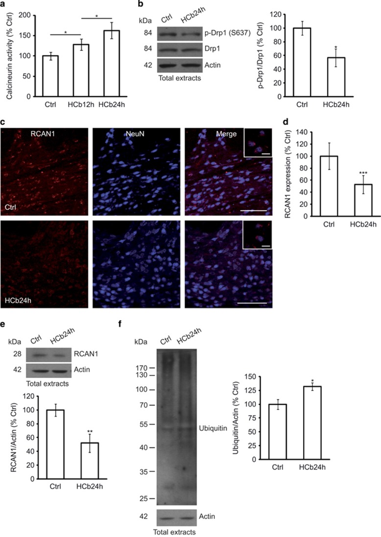
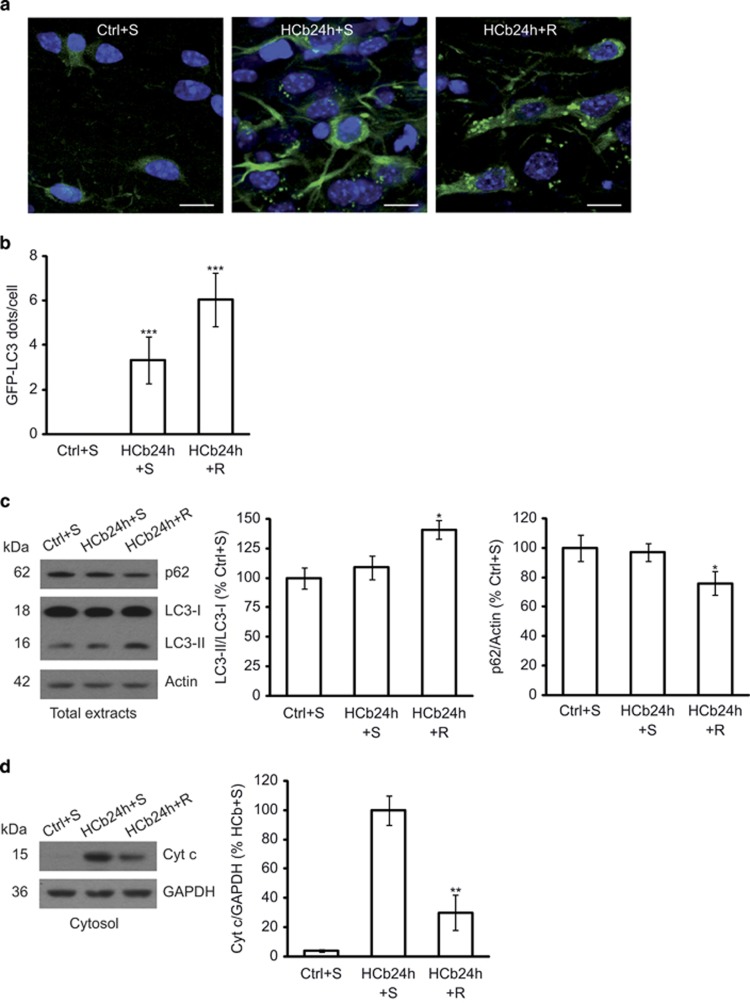
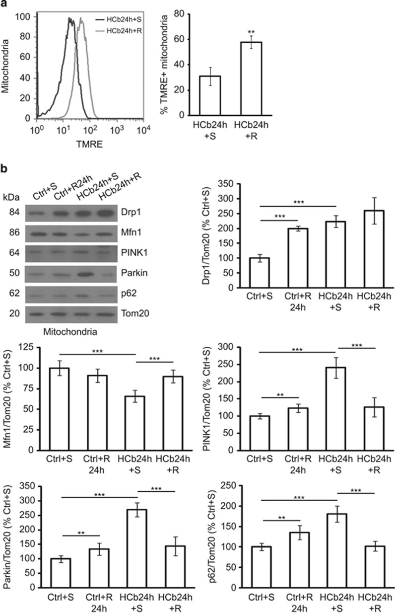
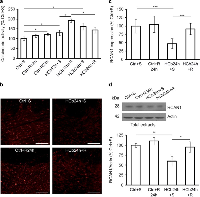
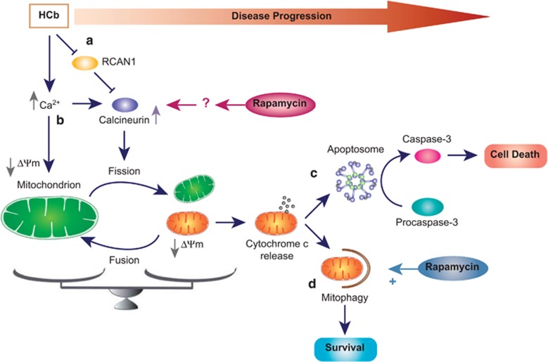
Mitochondria are key organelles for the maintenance of life and death of the cell, and their morphology is controlled by continual and balanced fission and fusion dynamics. A balance between these events is mandatory for normal mitochondrial and neuronal function, and emerging evidence indicates that mitochondria undergo extensive fission at an early stage during programmed cell death in several neurodegenerative diseases. A pathway for selective degradation of damaged mitochondria by autophagy, known as mitophagy, has been described, and is of particular importance to sustain neuronal viability. In the present work, we analyzed the effect of autophagy stimulation on mitochondrial function and dynamics in a model of remote degeneration after focal cerebellar lesion. We provided evidence that lesion of a cerebellar hemisphere causes mitochondria depolarization in axotomized precerebellar neurons associated with PTEN-induced putative kinase 1 accumulation and Parkin translocation to mitochondria, block of mitochondrial fusion by Mfn1 degradation, increase of calcineurin activity and dynamin-related protein 1 translocation to mitochondria, and consequent mitochondrial fission. Here we suggest that the observed neuroprotective effect of rapamycin is the result of a dual role: (1) stimulation of autophagy leading to damaged mitochondria removal and (2) enhancement of mitochondria fission to allow their elimination by mitophagy. The involvement of mitochondrial dynamics and mitophagy in brain injury, especially in the context of remote degeneration after acute focal brain damage, has not yet been investigated, and these findings may offer new target for therapeutic intervention to improve functional outcomes following acute brain damage.


Figures

Figure 1
Figure 1
HCb causes mitochondrial membrane depolarization and fragmentation in axotomized precerebellar nuclei. (a) Representative cytofluorimetric analysis (left panel) and graph showing quantitative analysis (right panel) of mitochondria isolated from precerebellar nuclei of Ctrl and lesioned mice at 24 h (HCb24h) and stained with tetramethylrhodamine ethyl ester (TMRE). Data are expressed as mean±S.D. (**P<0.01, n=4). (b) Representative transmission electron microscopy of precerebellar neurons in Ctrl and HCb mice at 24 h (HCb24h). (II and III) are higher magnification of a mitochondrion (m) shown in I. The cristae are well preserved and the electron density of mitochondrial matrix appears as regular. By contrast, the mitochondrion shown in I' and enlarged in II' and III' is profoundly altered, showing disrupted cristae and abnormal matrix. N, nucleus; ly, lysosomal vesicle. Scale bars, 1 μm (I and II) and 200 nm (III). (c) Representative immunoblots and densitometric graphs of mitochondrial content of Drp1, Mfn1, PINK1, Parkin and p62 levels (Ctrl is indicated as 100%) in precerebellar nuclei of Ctrl and HCb24h mice. The mitochondrial protein Tom20 was used as loading control. Data are expressed as mean±S.D. (**P<0.01, n=5)
Figure 2
Figure 2
Drp1 translocation to mitochondria in axotomized neurons is associated with increased calcineurin activity, RCAN1 downregulation, and protein ubiquitination. (a) Histogram showing calcineurin activity obtained by monitoring 32P release from radiolabeled purified RII substrate in axotomized precerebellar nuclei of Ctrl and HCb mice at 12 and 24 h (HCb12h and HCb24h, respectively). Data are expressed as mean (Ctrl percentage)±S.D. (*P<0.05, n=5). (b) Representative immunoblots and densitometric graphs of p-Drp1(S637)/Drp1 ratio (Ctrl percentage) in precerebellar nuclei of Ctrl and HCb24h mice. Actin was used as loading control. Data are expressed as mean±S.D. (*P<0.05, n=5) (c) Double-labeled and merged confocal images of RCAN1 (red) and NeuN (blue) in precerebellar nuclei of Ctrl and HCb mice at 24 h. Scale bars, 100 and 20 μm (inset). (d) Densitometric graph of RCAN1 levels in Ctrl and HCb24h mice. Data are expressed as mean±S.D. (***P<0.001, n=5 mice per group, N=150 cells per group). (e) Representative immunoblots and densitometric graphs of RCAN1 levels (Ctrl percentage) in precerebellar nuclei of Ctrl and HCb24h mice. Actin was used as loading control. Data are expressed as mean±S.D. (**P<0.01, n=5). (f) Representative immunoblots and densitometric analysis of proteins extracted from precerebellar nuclei of Ctrl and HCb24h mice probed with anti-multiubiquitin antibody. Actin was used as loading control. Data are expressed as mean±S.D. (*P<0.05, n=5). Ctrl is defined as 100%
Figure 3
Figure 3
Rapamycin treatment stimulates autophagy in HCb mice at 24 h and reduces cytochrome c release into the cytosol. (a) Double-labeled and merged confocal images of GFP-LC3 (green) and DAPI (blue) of precerebellar neurons in saline-treated Ctrl GFP-LC3 (Ctrl+S), saline-treated HCb GFP-LC3 (HCb24h+S), and rapamycin-treated HCb GFP-LC3 (HCb24h+R) mice at 24 h. Scale bars, 10 μm. (b) Histogram of the number of GFP-LC3 dots per neuron. Data are expressed as mean±S.D. (***P<0.001, n=5 mice per group, N=150 cells per group). (c) Representative immunoblots and densitometric graph of LC3-II/LC3-I ratio and p62 levels in precerebellar nuclei of saline-injected Ctrl (Ctrl+S), saline-injected HCb24h (HCb24h+S), and rapamycin-injected HCb24h (HCb24h+R) mice. Actin was used as loading control. Data are expressed as mean±S.D. (*P<0.05, n=5). (d) Representative immunoblots and densitometric graph of cytosolic proteins purified from precerebellar nuclei of Ctrl+S, HCb24h+S, and HCb24h+R mice. Cytosolic protein glyceraldehyde 3-phosphate dehydrogenase (GAPDH) was used as loading control. Data are expressed as mean±S.D. (**P<0.01, n=5). HCb+S is defined as 100%
Figure 4
Figure 4
Rapamycin treatment improves mitochondrial function and perturbs mitochondria dynamics. (a) Representative cytofluorimetric analysis (left) and quantification graphs (right) of mitochondria isolated from precerebellar nuclei of saline- and rapamycin-treated HCb (HCb24h+S and HCb24h+R, respectively) mice at 24 h and stained with tetramethylrhodamine ethyl ester (TMRE). Data are expressed as mean ±S.D. (**P<0.01, n=4). (b) Representative immunoblots and densitometric graph of mitochondrial levels of Drp, Mfn1, PINK1, Parkin, and p62 in precerebellar nuclei of saline- and rapamycin-treated Ctrl (Ctrl+S and Ctrl+R24h, respectively) and saline- and rapamycin-treated HCb (HCb24h+S and HCb24h+R, respectively) mice. The mitochondrial protein Tom20 was used as loading control. Data are expressed as mean±S.D. (**P<0.01, ***P<0.001, n=7). Ctrl+S is indicated as 100%
Figure 5
Figure 5
Rapamycin treatment stimulates calcineurin activity. (a) Histogram of calcineurin activity obtained by monitoring 32P release from radiolabeled purified RII substrate in axotomized precerebellar nuclei of saline- and rapamycin-treated Ctrl (Ctrl+S, Ctrl+R12h, and Ctrl+R24h) and saline- and rapamycin-treated HCb (HCb12h+S, HCb12h+R, HCb24h+S, and HCb24h+R) mice at 12 and 24 h. Data are expressed as mean (Ctrl+S percentage)±S.D. (*P<0.05, n=5). (b) Confocal images of RCAN1 staining of precerebellar nuclei of Ctrl+S, Ctrl+R, HCb+S, and HCb+R mice at 24 h. (c) Densitometric graph of RCAN1 staining in Ctrl+S, Ctrl+R24h, HCb24h+S, and HCb24h+R mice. Data are expressed as mean±S.D. (***P<0.001, n=6 mice per group, N=180 cells per group). (d) Representative immunoblots and densitometric graph of RCAN1 levels (Ctrl+S percentage) in precerebellar nuclei of Ctrl+S, Ctrl+R24h, HCb24h+S, and HCb24h+R mice at 24 h. Actin was used as loading control. Data are expressed as mean ±S.D. (*P<0.05, **P<0.01, n=5)
Figure 6
Figure 6
Schematic of the neuroprotective effect of rapamycin in hemicerebelloctomized mice. HCb causes a reduction in levels of the calcineurin inhibitor RCAN1, leading to an increase in calcineurin activity in precerebellar nuclei contralateral to the lesion (a). Concomitant with the RCAN1 degradation, the axotomy-induced increase in intracellular Ca2+, on the one hand enhances calcineurin activity and on the other hand affects the mitochondrial membrane potential (ΔΨm) (b). Calcineurin activity promotes mitochondrial fission inducing Drp1 translocation (see text). Damaged mitochondria release cytochrome c into the cytosol, activating the apoptotic pathway, leading to cell death (in absence of mitophagy) (c). Prompt activation of the autophagic pathway might help remove damaged organelles by mitophagy, promoting neuronal survival (d). In this model, rapamycin treatment has two functions: (1) stimulate autophagy, leading to removal of damaged mitochondria; and (2) enhance mitochondrial fission to effect their elimination by mitophagy

References

    1. Kroemer G, Dallaporta B, Resche-Rigon M. The mitochondrial death/life regulator in apoptosis and necrosis. Annu Rev Physiol. 1998;60:619–642. - PubMed
    1. Hoppins S, Lackner L, Nunnari J. The machines that divide and fuse mitochondria. Annu Rev Biochem. 2007;76:751–780. - PubMed
    1. Chen H, Chan DC. Physiological functions of mitochondrial fusion. Ann N Y Acad Sci. 2010;1201:21–25. - PubMed
    1. Chang CR, Blackstone C. Dynamic regulation of mitochondrial fission through modification of the dynamin-related protein Drp1. Ann N Y Acad Sci. 2010;1201:34–39. - PMC - PubMed
    1. James DI, Parone PA, Mattenberger Y, Martinou JC. hFis1, a novel component of the mammalian mitochondrial fission machinery. J Biol Chem. 2003;278:36373–36379. - PubMed

Publication types

MeSH terms