. 2015 Jan 16;290(3):1592-606.
doi: 10.1074/jbc.M114.588590. Epub 2014 Nov 27.

Phospholipid-binding sites of phosphatase and tensin homolog (PTEN): exploring the mechanism of phosphatidylinositol 4,5-bisphosphate activation

Affiliations

Phospholipid-binding sites of phosphatase and tensin homolog (PTEN): exploring the mechanism of phosphatidylinositol 4,5-bisphosphate activation

Yang Wei et al. J Biol Chem. .

Abstract

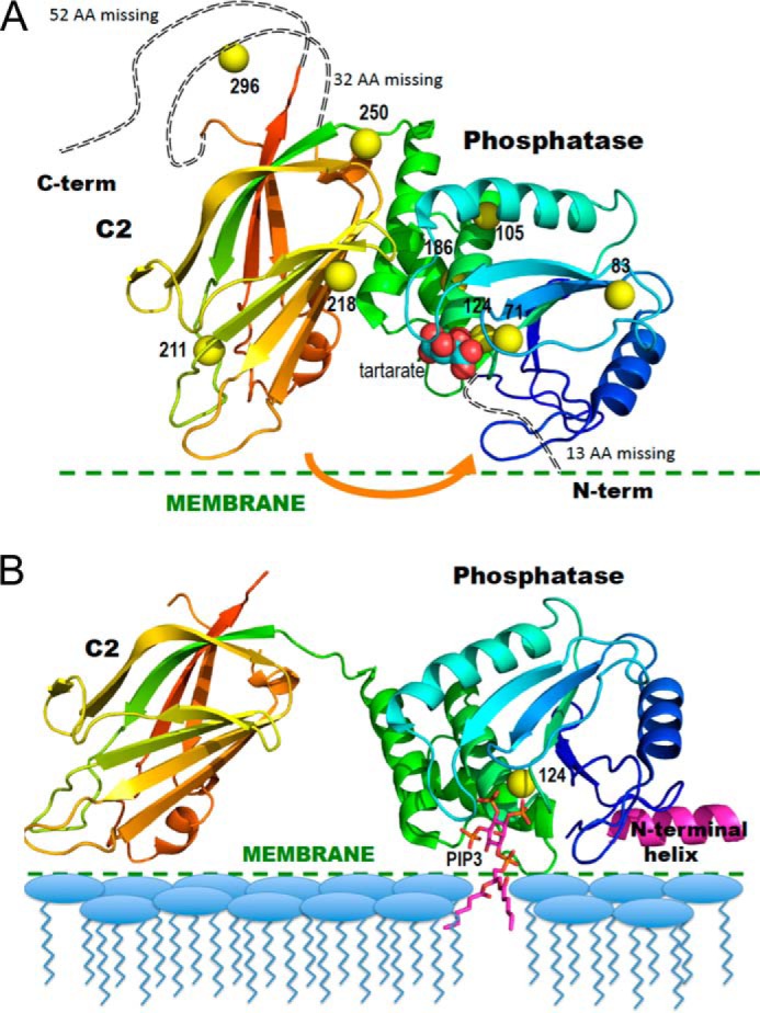
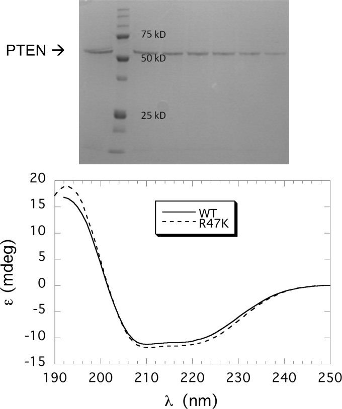
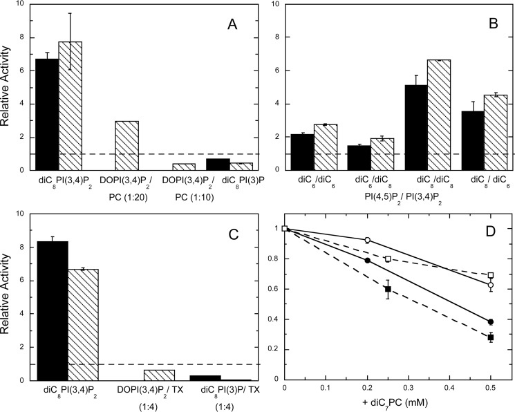
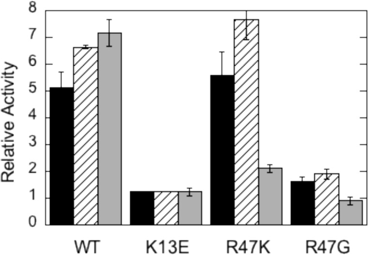
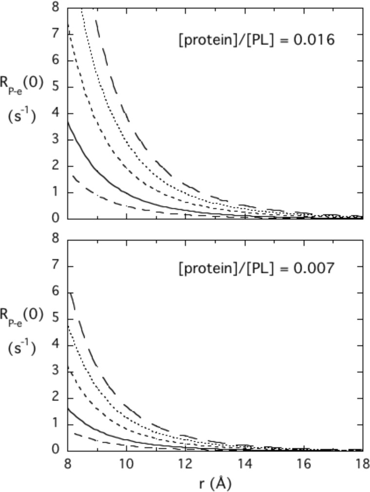
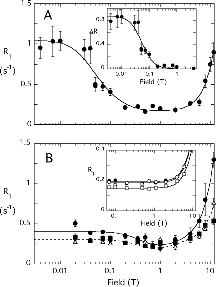
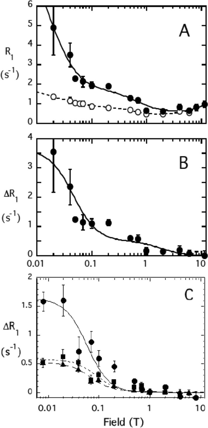
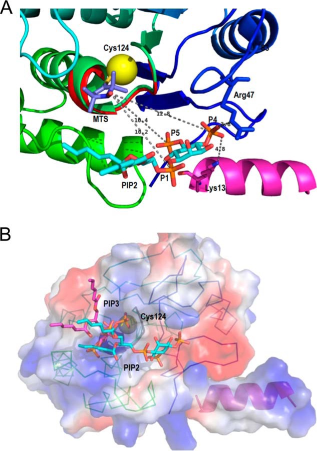
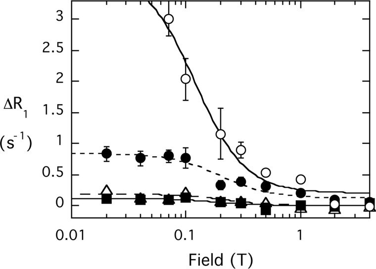
The lipid phosphatase activity of the tumor suppressor phosphatase and tensin homolog (PTEN) is enhanced by the presence of its biological product, phosphatidylinositol 4,5-bisphosphate (PI(4,5)P2). This enhancement is suggested to occur via the product binding to the N-terminal region of the protein. PTEN effects on short-chain phosphoinositide (31)P linewidths and on the full field dependence of the spin-lattice relaxation rate (measured by high resolution field cycling (31)P NMR using spin-labeled protein) are combined with enzyme kinetics with the same short-chain phospholipids to characterize where PI(4,5)P2 binds on the protein. The results are used to model a discrete site for a PI(4,5)P2 molecule close to, but distinct from, the active site of PTEN. This PI(4,5)P2 site uses Arg-47 and Lys-13 as phosphate ligands, explaining why PTEN R47G and K13E can no longer be activated by that phosphoinositide. Placing a PI(4,5)P2 near the substrate site allows for proper orientation of the enzyme on interfaces and should facilitate processive catalysis.

Keywords: 31P Field Cycling; Enzyme Kinetics; Micelles; Nuclear Magnetic Resonance (NMR); Phosphatase and Tensin Homolog (PTEN); Phosphatidylinositol Phosphatase; Phosphoinositide; Phospholipid-binding Site; Spin-labeled Protein.


Figures

FIGURE 1.
FIGURE 1.
Ribbon diagrams of the crystal structure of PTEN in the absence and presence of substrate-containing membrane. A, PTEN crystal structure (Protein Data Bank code 1D5R) with colors from blue to red for N to C termini. Yellow spheres represent sulfur atoms of Cys residues that are spin-labeled in the NMR experiments. The tartrate molecule bound at the postulated active site is in van der Waals sphere representation. The arrow indicates a postulated movement of the two domains upon membrane binding. B, model for membrane-bound PTEN with the missing N-terminal peptide docked as an interfacial helix (pink). The sulfur of Cys-124 is depicted as a yellow sphere and a diC6PIP3 molecule (magenta) is docked in the active site. The postulated conformational change is based on that suggested by Kalli et al. (17).
FIGURE 2.
FIGURE 2.
Purification of recombinant PTEN and variants. Top, SDS-PAGE analysis of fractions from the Ni-NTA column; mass standards are indicated on the gel. Bottom, CD spectra for recombinant PTEN (–) and R47A (- - -) are indicated.
FIGURE 3.
FIGURE 3.
Effect of PI(4,5)P2 and other amphiphiles on PTEN hydrolysis of different substrates. A, relative specific activities for PTEN toward 0.1 mm diC8PI(3,4)P2, DOPI(3,4)P2/POPC (0.05 mm/0.95 mm) LUVs, DOPI(3,4)P2/POPC (0.1 mm/0.9 mm) LUVs, and 0.5 mm diC8PI(3)P with PI(4,5)P2 are shown, with substrate/PI(4,5)P2 = 0.5 (■) or 1.0 (\\\\). B, effect of varying the chain length of substrate and activator. Substrates include 0.1 mm diC6PI(3,4)P2 or 0.1 mm diC8PI(3,4)P2 with dihexanoyl- or dioctanoyl-PI(4,5)P2 added, and substrate/PI(4,5)P2 = 0.5 (■) or 1.0 (\\\\). C, effect of PIP2 and Triton X-100 on PTEN hydrolysis of different substrates. Substrates include diC8PI(3,4)P2/Triton X-100 (0.1 to 0.4 mm), DOPI(3,4)P2/Triton X-100 (0.1 to 0.4 mm), and diC8PI(3)P/Triton X-100 (0.5 to 2 mm). The concentration of PI(4,5)P2 (either the dioctanoyl compound or DOPI(4,5)P2 depending on the substrate) was half (■) or the same (\\\\) as that of the substrate. The dashed lines in A–C would represent an activity with the PI(4,5)P2 equivalent to that in its absence. D, diC7PC inhibition of PTEN activity toward 0.5 mm diC8PI(3)P (■, □) or 0.1 mm diC8PI(3,4)P2 (●, ○) in the absence (filled symbols) or presence (open symbols) of Triton X-100 equivalent to four times the substrate concentration. Error bars here (and in other plots of enzyme activities) represent standard deviations from the repeats for each experimental condition.
FIGURE 4.
FIGURE 4.
Effect of PI(4,5)P2 on the activity of recombinant PTEN and variants K13E, R47K, and R47G in monomer and vesicle assay systems. The ratio of the specific activity with PI(4,5)P2 added to the activity in its absence is shown for WT and the three mutant proteins. In the monomer substrate mixture diC8PI(3,4)P2 is 0.1 mm with either 0.05 mm (■) or 0.1 mm (///) diC8PI(4,5)P2. Vesicles (▩) were composed of DOPI(3,4)P2/POPC (0.05 to 0.95 mm) with 0.05 mm DOPI(4,5)P2. The specific activities of wild type PTEN toward each substrate in the absence of PIP2 are 45 ± 4 nmol/mg min for diC8PI(3,4)P2 and 0.59 ± 0.02 nmol/mg min for DOPI(3,4)P2/POPC LUVs.
FIGURE 5.
FIGURE 5.
Dependence of RP-e(0) on the distance of a 31P nucleus from an unpaired electron and the τP-e correlation time predicted for two different ratios of protein to the 31P-containing amphiphile. Different τP-e values are 25 (– – –), 50 (—), 100 (- - -), 150 (···), and 200 (— — —) ns.
FIGURE 6.
FIGURE 6.
Field dependence of the PR1E for diC6PI and diC6PI(4,5)P2. A, R1 for the 31P of diC6PI (3 mm) caused by 4.9 μm spin-labeled PTEN; the inset shows the ΔR1 of this phosphodiester from 0.004 to 4 T after subtraction of the profile obtained using nonlabeled PTEN. B, R1 for the 31P resonances of diC6PI(4,5)P2 (3 mm) in the presence of 5.1 μm spin-labeled PTEN: P-1 (●), P-4 (▵), and P-5 (▴). The inset in B shows the R1 profile for 3 mm I(1,4,5)3 in the presence of 4.9 μm spin-labeled PTEN: P-4 (●), P-1 phosphate (○), and P-5 phosphate (□). Note that none of these 31P resonances exhibit the low field rise in R1 indicative of a small molecule-protein complex. Error bars for R1 values (here and in subsequent plots) were obtained from a least squares fit of the ln(intensity) versus time determining R1 at that particular field.
FIGURE 7.
FIGURE 7.
Field dependence of the PR1E for diC8PI and diC8PI(4,5)P2. A, R1 for the 31P of diC8PI (3.0 mm) in the presence of 2.0 μm unlabeled (○) or spin-labeled (●) PTEN is shown as a function of magnetic field. B, ΔR1 representing the difference due to the spin-labeled PTEN is shown; the line represents a fit with two dispersions with τP-e values of 5 and 200 ns. C, ΔR1 for diC8PI(4,5)P2 (3.0 mm) in the presence of 4.9 μm spin-labeled PTEN (solid symbols): P-1 (●), P-4 (▴), and P-5 (■).
FIGURE 8.
FIGURE 8.
Field dependence of the 31P PR1E for diC8PI and diC8PI(4,5)P2 in the presence of spin-labeled PTEN C124S. A, effect of 4.3 μm spin-labeled C124S on 3 mm diC8PI(4,5)P2: P-1 (●); P-4 (□); (▴) P-5. B, comparison of PRE for diC8PI (3 mm) produced by spin-labeled 2.0 μm native PTEN (○) or 2.2 μm spin-labeled variant C124S (●); the field dependence was fit with τP-e = 200 ns, and the arrow approximates the amount of R1 contributed by a spin label on Cys-124.
FIGURE 9.
FIGURE 9.
Ribbon diagrams of the model of diC6PI(4,5)P2 binding to PTEN at a discrete site near the active site are compatible with the 31P electron distances to the Cys-124 spin label. A, view of the phosphatase domain as seen from the membrane. The sulfur of Cys-124 is the yellow sphere to which the 2,2,5,5-tetramethyl-1-oxyl-3-methyl methanethiosulfonate is attached. The PI(4,5)P2 molecule, placed according to the surface features and to the relative distances determined by NMR, forms hydrogen-bonded contacts with Arg-47 and Lys-13, both critical residues for the PIP2 activation to be observed. B, surface representation of the phosphatase domain, in a similar view to that in A, colored by the electrostatic potential (red, negative, and blue, positive) with both substrate and activator docked to their sites. The PI(3,4,5)P3 molecule (magenta) is docked at the active site marked by the dark loop and the yellow sphere depicting Cys-124. The PI(4,5)P2 molecule (in cyan) occupies the same site as in A. The N-terminal helix is docked into the phosphatase domain, and its positive electrostatic potential complements the charges of the domain to create a positive membrane binding profile.
FIGURE 10.
FIGURE 10.
Effect of spin-labeled K13E PTEN (4.1 μm) on the field dependence of R1 for phospholipid mixed micelles of 3 mm diC8PI () and 3 mm diC8PI(4,5)P2 (P-1 (), P-4, (Δ), P-5 ()).

References

    1. Song M. S., Salmena L., Pandolfi P. P. (2012) The functions and regulation of the PTEN tumour suppressor. Nat. Rev. Mol. Cell Biol. 13, 283–296 - PubMed
    1. Salmena L., Carracedo A., Pandolfi P. P. (2008) Tenets of PTEN tumor suppression. Cell 133, 403–414 - PubMed
    1. Leslie N. R., Downes C. P. (2004) PTEN function: how normal cells control it and tumour cells lose it. Biochem. J. 382, 1–11 - PMC - PubMed
    1. Chang N., El-Hayek Y. H., Gomez E., Wan Q. (2007) Phosphatase PTEN in neuronal injury and brain disorders. Trends Neurosci. 30, 581–586 - PubMed
    1. Maehama T., Dixon J. E. (1998) The tumor suppressor, PTEN/MMAC1, dephosphorylates the lipid second messenger, phosphatidylinositol 3,4,5-trisphosphate. J. Biol. Chem. 273, 13375–13378 - PubMed

Publication types

Associated data