. 2014 Nov 28;16(6):488.
doi: 10.1186/s13075-014-0488-y.

Intra-articular injection of synthetic microRNA-210 accelerates avascular meniscal healing in rat medial meniscal injured model

Intra-articular injection of synthetic microRNA-210 accelerates avascular meniscal healing in rat medial meniscal injured model

Yoshitaka Kawanishi et al. Arthritis Res Ther. .

Abstract

Introduction: The important functions of the meniscus are shock absorption, passive stabilization and load transmission of the knee. Because of the avascularity of two-thirds of the meniscal center region, the treatment of tears in this area is hard. Recently, microRNAs have been proven to play an important role in the pathogenesis of diseases. We focused on microRNA (miR)-210, which plays a wide spectrum of roles comprising mitochondrial metabolism, angiogenesis, DNA repair and cell survival. This study aimed to investigate the effect of intra-articular injection of synthetic miR-210 on the injured meniscus in the avascular zone.

Methods: The middle segments of the medial meniscus of Spraque Dawley rats were incised longitudinally with a scalpel. An intra-articular injection of double-stranded (ds) miR-210 (for control group using control dsRNA) with atelocollagen was administered immediately after injury. Four weeks and 12 weeks after the injection, we conducted a histologic evaluation, immunohistochemical evaluation and Real-time PCR analysis. In vitro, the inner meniscus and synovial cells were isolated from rat knee joint, and were transfected with ds miR-210 or control dsRNA. Real-time PCR and immunohistochemical evaluations were performed.

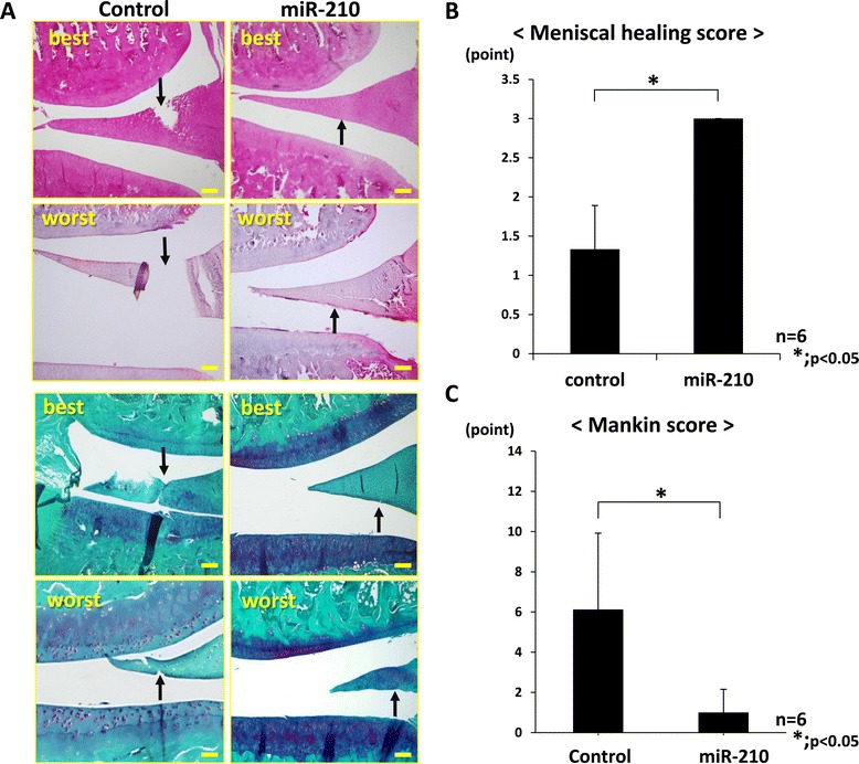
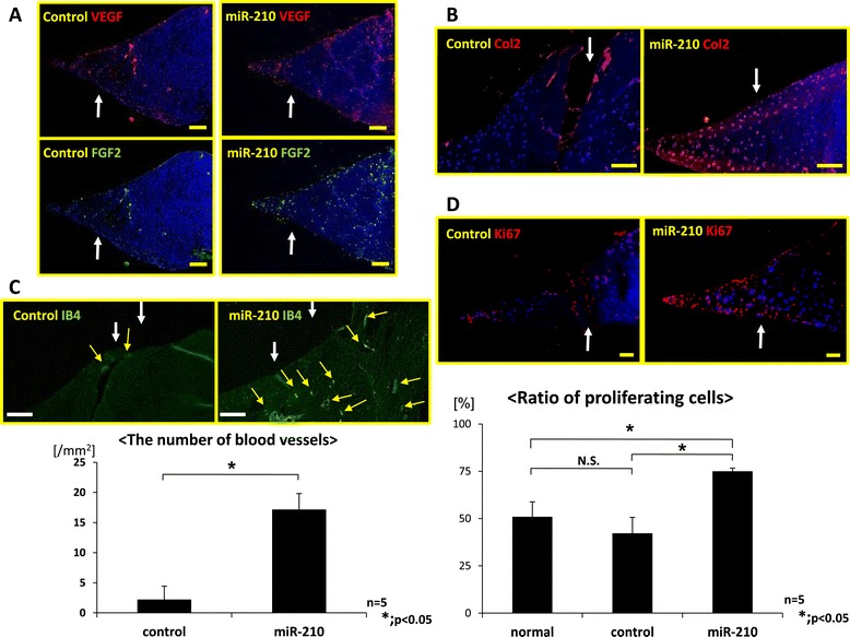
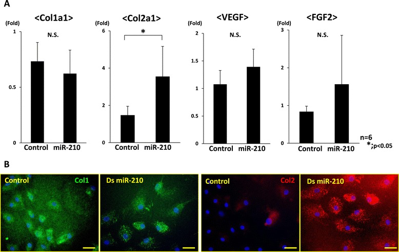
Results: Twenty-four hours after the injection, FAM (Fluorescein amidite) labeled miR-210 was observed in the cells around the injured site. Four weeks after the injection, the injured site of the miR-210 group was filled with repaired tissue while that of the control was not repaired. In gene expression analysis of the meniscus, the expression of miR-210, Collagen type 2 alpha 1 (Col2a1), Vascular endothelial growth factor (VEGF), and Fibroblast growth factor-2 (FGF2) in the miR-210 group was significantly higher than that in the control. At 12 weeks, the intra-articular injection of miR-210 had healed the injured site of the meniscus and had prevented articular cartilage degeneration. In vitro, miR-210 upregulated Col2a1 expression in the meniscus cells and VEGF and FGF2 expression in the synovial cells.

Conclusions: An intra-articular injection of ds miR-210 was effective in the healing of the damaged white zone meniscus through promotion of the collagen type 2 production from meniscus cells and through upregulated of VEGF and FGF2 from synovial cells.


Figures

Figure 1
Figure 1
Histological findings of the meniscus at 4 weeks. (A) Hematoxylin and eosin staining. (B) Safranin O staining: the injured site of the miR-210 group was filled with repaired tissue stained with safranin O, while that of the control group was not repaired. Upper panels, best result; lower panels, worst result (bar = 100 μm). (C) Histological score of the miR-210 group (2.3 ± 0.8 points) was significantly higher than that of the control group (1.0 ± 1.0 point) (*P < 0.05, n = 6 for each group). (D) Detection of fluorescein amidite (FAM)-labeled double-stranded (ds) miR-210: distribution of green fluorescence was observed in the meniscus around the injured site 24 hours after injection of the FAM-labeled ds miR-210/atelocollagen complex into the joint (bar = 100 μm). Expression of green fluorescence was observed in the cytoplasm of cells in high-power views (bar = 25 μm). Arrows, FAM-labeled ds miR-210/atelocollagen complex. DAPI, 4′,6-diamidino-2-phenylindole.
Figure 2
Figure 2
Histological findings of the knee joint at 12 weeks. (A) Hematoxylin and eosin staining and safranin O staining: the injured site in the miR-210 group was morphologically healed whereas the tear in the meniscus still remained in the control group (bar = 100 μm). Safranin O staining revealed that the degeneration of articular cartilage in the miR-210 group was lower than that of the control group. Arrow, injured site. Upper panels, best result; lower panels, worst result. (B) Meniscal healing score in the miR-210 group (3.0 ± 0.0 points) was significantly higher than in the control group (1.3 ± 0.6 points) (*P < 0.05, n = 6 for each group). Degenerative change of cartilage in the control group was more advanced than in the miR-210 group. (C) Mankin score in the miR-210 group (1.0 ± 0.4 points) was significantly lower than in the control group (5.6 ± 1.4 points), showing a significant difference between the two groups (*P < 0.05, n = 6 for each group). Intra-articular injection of miR-210 prevented articular cartilage degeneration.
Figure 3
Figure 3
Gene expression analysis by real-time PCR in the meniscus at 4 weeks following intraarticular injection. Expression of mature miR-210, collagen type 1 alpha 1 (Col1a1), collagen type 2 alpha 1 (Col2a1), vascular endothelial growth factor (VEGF) and fibroblast growth factor-2 (FGF2) was examined using real-time PCR. Expression of miR-210, Col2a1, VEGF and FGF2 in the miR-210 group was significantly higher than that in the control and normal groups (*P < 0.05). There was no significant difference in miR-210, Col2a1, VEGF and FGF2 expression between the control and normal groups (*P < 0.05, N.S. (no significant difference), n = 5 for each group).
Figure 4
Figure 4
Immunohistochemistry of meniscus at 4 weeks after intra-articular injection. (A) Immunohistochemistry of vascular endothelial growth factor (VEGF; upper) and fibroblast growth factor-2 (FGF2; lower). VEGF and FGF2 expressed intensely on the surface of the meniscus, around the injured site and in the red zone in the miR-210 group compared with the control group. Arrow, injured site. Bar = 100 μm. (B) Immunohistochemistry of type 2 collagen. Type 2 collagen expression was observed around the injured site of the meniscus in the miR-210 group compared with the control group. Arrow, injured site. Bar = 100 μm. (C) Isolectin B4 staining and the number of blood vessels. Newly formed vessels were observed around the injured site in the miR-210 group, while little blood vessels were observed in the control. White arrow, injured site; yellow arrow, blood vessels. Bar = 100 μm. Number of blood vessels in the miR-210 group was significantly higher than that in the control group (*P < 0.05, n = 5 for each group). (D) Immunohistochemistry of Ki67 and the ration of proliferative cells. In the miR-210 group, many proliferative cells were observed around the injured site. Arrow, injured site. Proliferative cells in the miR-210 group were significantly higher than that in the control group (*P < 0.05, N.S. (no significant difference), n = 5 for each group).
Figure 5
Figure 5
Gene expression analyses in inner meniscus cells after overexpression of miR-210. (A) Real-time PCR analysis of collagen type 1 alpha 1 (Col1a1), collagen type 2 alpha 1 (Col2a1), vascular endothelial growth factor (VEGF) and fibroblast growth factor-2 (FGF2) at 7 days after in vitro transfection of inner meniscus cells. Expression of only Col2a1 was significantly higher than that in the control group (*P < 0.05, N.S. (no significant difference), n = 6 for each group). (B) Immunohistochemical analysis indicates that collagen type 2 was highly expressed in the miR-210 group (bar = 100 μm). These results demonstrate that miR-210 could enhance the expression of collagen type 2 in the meniscus cells. ds, double stranded.
Figure 6
Figure 6
Gene expression analyses in synovial cells after overexpression of miR-210. (A) Real-time PCR analysis of collagen type 1 alpha 1 (Col1a1), collagen type 2 alpha 1 (Col2a1), vascular endothelial growth factor (VEGF) and fibroblast growth factor-2 (FGF2) at 7 days after in vitro transfection of synovial cells. Expression of Col2a1 was not detected in both groups. Expression of VEGF and FGF2 was significantly higher than in the control group (*P < 0.05, N.S. (no significant difference), n = 6 for each group). (B) Immunohistochemical analysis indicates that VEGF and FGF2 were highly expressed in the miR-210 group (bar = 100 μm). These results demonstrated that miR-210 could enhance the expression of VEGF and FGF2 expression in synovial cells. ds, double stranded.

References

    1. Bullough PG, Munuera L, Murphy J, Weinstein AM. The strength of the menisci of the knee as it relates to their fine structure. J Bone Joint Surg Br. 1970;52:564–567. - PubMed
    1. Petersen W, Tillmann B. Collagenous fibril texture of the human knee joint menisci. Anat Embryol. 1998;197:317–324. doi: 10.1007/s004290050141. - DOI - PubMed
    1. Arnoczky SP, Warren RF. Microvasculature of the human meniscus. Am J Sports Med. 1982;10:90–95. doi: 10.1177/036354658201000205. - DOI - PubMed
    1. Petersen W, Pufe T, Starke C, Fuchs T, Kopf S, Raschke M, Becker R, Tillmann B. Locally applied angiogenic factors – a new therapeutic tool for meniscal repair. Ann Anat. 2005;187:509–519. doi: 10.1016/j.aanat.2005.04.010. - DOI - PubMed
    1. Englund M, Roos EM, Lohmander LS. Impact of type of meniscal tear on radiographic and symptomatic knee osteoarthritis: a sixteen-year followup of meniscectomy with matched controls. Arthritis Rheum. 2003;48:2178–2187. doi: 10.1002/art.11088. - DOI - PubMed

Publication types

LinkOut - more resources