. 2015 Feb;95(4):660-77.
doi: 10.1111/mmi.12893. Epub 2014 Dec 30.

Enterococcus faecalis pCF10-encoded surface proteins PrgA, PrgB (aggregation substance) and PrgC contribute to plasmid transfer, biofilm formation and virulence

Affiliations

Enterococcus faecalis pCF10-encoded surface proteins PrgA, PrgB (aggregation substance) and PrgC contribute to plasmid transfer, biofilm formation and virulence

Minny Bhatty et al. Mol Microbiol. 2015 Feb.

Abstract

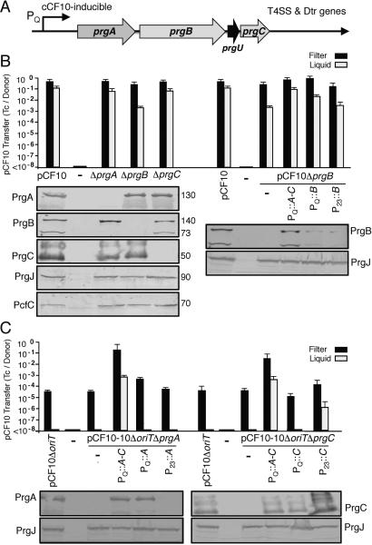
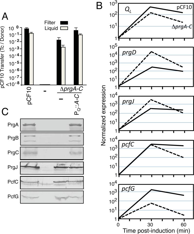
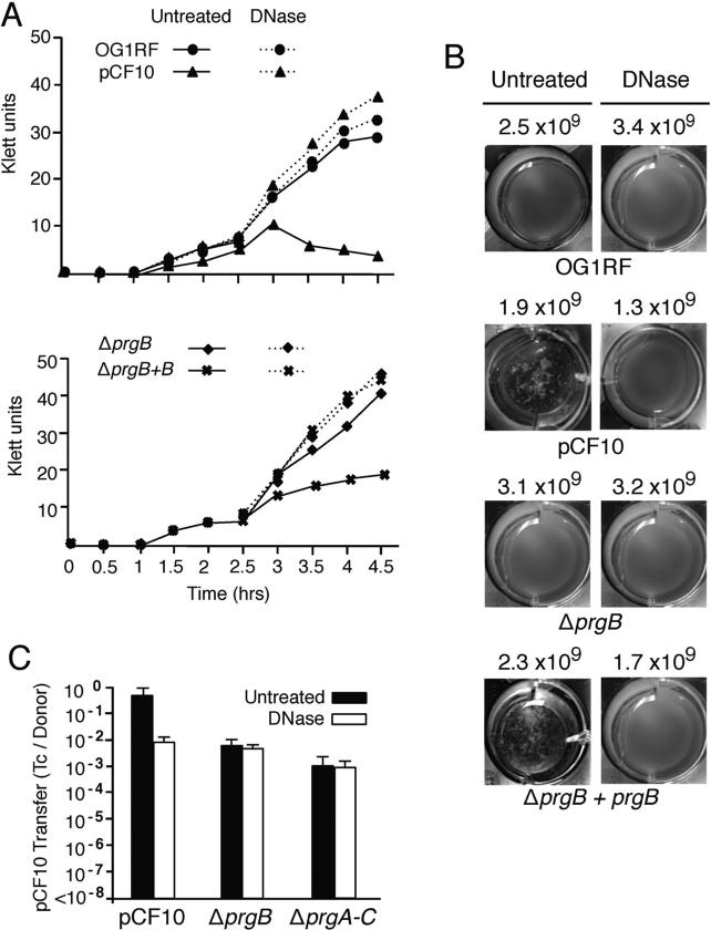
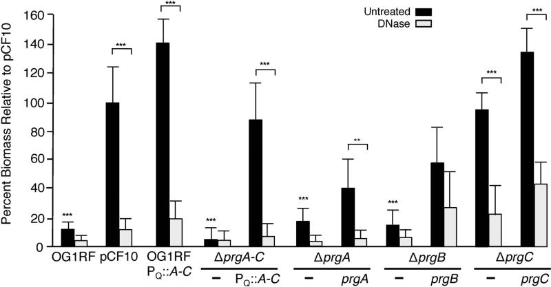
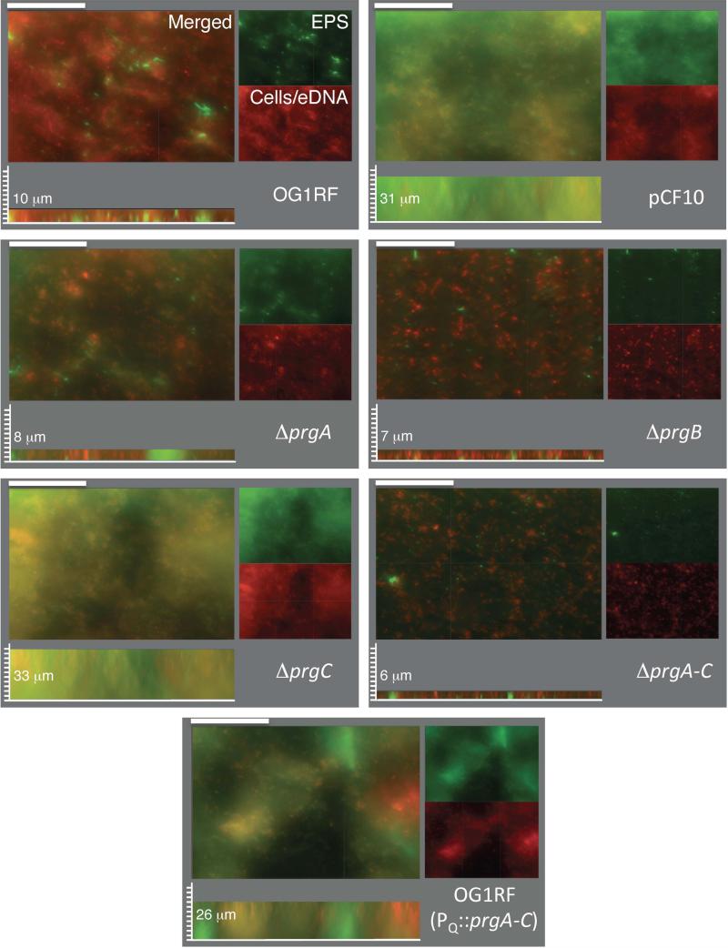
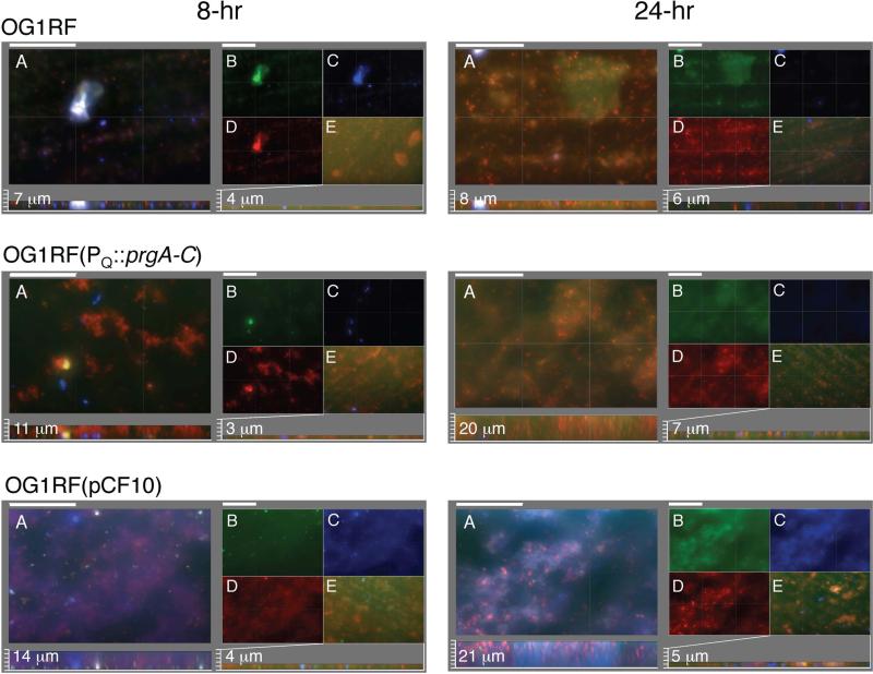
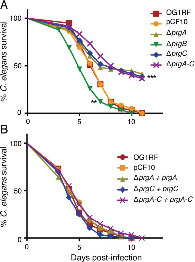
Enterococcus faecalis pCF10 transfers at high frequencies upon pheromone induction of the prgQ transfer operon. This operon codes for three cell wall-anchored proteins - PrgA, PrgB (aggregation substance) and PrgC - and a type IV secretion system through which the plasmid is delivered to recipient cells. Here, we defined the contributions of the Prg surface proteins to plasmid transfer, biofilm formation and virulence using the Caenorhabditis elegans infection model. We report that a combination of PrgB and extracellular DNA (eDNA), but not PrgA or PrgC, was required for extensive cellular aggregation and pCF10 transfer at wild-type frequencies. In addition to PrgB and eDNA, production of PrgA was necessary for extensive binding of enterococci to abiotic surfaces and development of robust biofilms. However, although PrgB is a known virulence factor in mammalian infection models, we determined that PrgA and PrgC, but not PrgB, were required for efficient killing in the worm infection model. We propose that the pheromone-responsive, conjugative plasmids of E. faecalis have retained Prg-like surface functions over evolutionary time for attachment, colonization and robust biofilm development. In natural settings, these biofilms are polymicrobial in composition and constitute optimal environments for signal exchange, mating pair formation and widespread lateral gene transfer.


Figures

FIG. 1
FIG. 1. Individual prg genes enhance but are not required for pCF10 transfer
(A) Schematic representation of the pCF10 prg gene cluster encoding PrgA (891 residues), PrgB (1,305 residues), PrgU (118 residues), PrgC (285 residues) relative to the pheromone-inducible PQ promoter and downstream T4SS and Dtr processing genes. (B) Histogram: Transfer frequencies of pCF10 plasmids in 2 h filter and liquid matings. Donor strains: OG1RF lacking () or carrying pCF10 or pCF10Δprg mutant plasmids (left), or pCF10ΔprgB and complementing plasmids expressing the prg genes (right). Complementing plasmids: PQ::prgA-C (pINY1801), PQ::prgB (pMB6), P23::prgB (pMB3). Transfer frequencies are presented as the number of transconjugants per donor cell (Tc's/Donor). Experiments were repeated at least 3 times and the histogram depicts the average values with standard deviations. Immunoblots: Steady state levels of PrgA, PrgB, PrgC,PrgJ and PcfC in E. faecalis OG1RF strains lacking (-) or carrying the pCF10 variants and prg expression constructs listed above each lane. The immunoblots were developed with antibodies to the proteins listed and the approximate size of the detected protein in kilodaltons (kDa's) is listed at the right. Protein extracts were loaded on a per-cell equivalent basis. (C) Histogram: Transfer frequencies in 2 h filter and liquid matings with OG1RF donors carrying pCF10ΔoriT, pCF10ΔoriTΔprgA, or pCF10ΔoriTΔprgC and complementing plasmids expressing the genes listed. Complementing plasmids: PQ::prgA-C (pINY1801), PQ::prgA (pMB5), P23::prgA (pMB2), PQ::prgC (pMB7), P23::prgC (pMB4). Immunoblots: Steady-state levels of PrgA, PrgC, or PrgJ in the strains listed above each lane as detected by Western immunoblotting.
FIG. 2
FIG. 2. Deletion of the entire prgA-C gene cluster only modestly diminishes plasmid transfer
(A) Transfer frequencies of OG1RF strains lacking or carrying pCF10, pCF10ΔprgA-C alone or with the PQ::prgA-C expression plasmid pINY1801 in 2 h filter and liquid matings. Transfer frequencies are presented as the number of transconjugants per donor cell (Tc's/Donor). Experiments were repeated at least 3 times and the histogram depicts average values with standard deviations. (B) qRT-PCR results showing the relative expression levels of regions of the prgQ operon located upstream (QL) and downstream (prgD, prgJ, pcfC, pcfG) of the prgA-C genes in pCF10 (solid lines) and pCF10ΔprgA-C (dashed lines) at 30 and 60 min following cCF10 pheromone induction with 5ng/ml cCF10. The data shown are from one biological replicate, which was repeated with similar results. (C) Steady state levels of pCF10-encoded surface proteins (PrgA, PrgB, PrgC) and downstream T4SS machine subunits (PrgJ, PcfC, PcfG) in E. faecalis OG1RF without (−) and with pCF10; pCF10ΔprgA-C or pCF10ΔprgA-C and the PQ::prgA-C expression plasmid pINY1801. Immunoblots were developed with antibodies to the proteins listed on the left. Protein extracts were loaded on a per-cell equivalent basis.
FIG. 3
FIG. 3. PrgB-mediated aggregation and plasmid transfer is dependent on eDNA
(A) Aggregation assays showing the effects of DNase treatment on pheromone (cCF10)-induced cellular aggregation. DNase treatment inhibited aggregation of OG1RF(pCF10) (upper graph) and OG1RF(pCF10ΔprgB, pMB6) expressing PQ::prgB (lower graph). (B) Photographs of cells grown in microtiter plates showing the effects of DNase on pheromone-induced clumping at 14 h following pheromone induction. Strains: OG1RF lacking or carrying pCF10, pCF10ΔprgB, or pCF10ΔprgB and pMB6. The numbers correspond to colony forming units (CFU's) per ml at 14 h following cCF10 induction in absence (Untreated) or presence of DNase. (C) Histograms depict the transfer frequencies of pCF10, pCF10ΔprgB and pCF10ΔprgA-C without or with DNase added at the onset of a 2 h liquid mating.
FIG. 4
FIG. 4. PrgA and PrgB mediate DNase-sensitive adherence to polystyrene
Biofilm formation by prg mutant strains on polystyrene microtiter plates. Strains: OG1RF, OG1RF(pCF10), or OG1RF strains carrying Δprg mutant plasmids and complementing plasmids are shown. Complementing plasmids: PQ::prgA (pMB5), PQ::prgB (pMB6), PQ::prgC (pMB7), and PQ::prgA-C (pINY1801). The biofilm biomass was assayed as a function of crystal violet stain retained. DNase treatment was initiated at the onset of cCF10 induction and biofilm formation and continued for the full incubation time of 24 h. Results are expressed as the percentage biomass relative to pCF10 and represent the average of at least three independent experiments; the error bars represent standard deviation. Statistically significant differences (***P<0.001; **P<0.01) were evaluated within each strain with respect to DNase treatment (brackets) and across the untreated group with respect to strains containing pCF10 (no brackets) using one-way analysis of variance followed by Newman-Keuls post hoc test.
FIG. 5
FIG. 5. PrgA and PrgB mediate development of robust, mature biofilms on PMMA coupons
Macroscopic images of biofilms produced by strains inoculated on PMMA coupons at the onset of cCF10 pheromone induction and incubated for 48 h in TSB medium. Cells and eDNA were stained with hexidium iodide (red), extracellular polysaccharide (EPS) was stained with calcofluor white (green). Merged two-color images and corresponding separated color channels of representative 48-h biofilms at 40× magnification are shown; scale bars, 50 μm. Sagittal views are shown depicting biofilm thicknesses with measured values shown in μM; scale bars, 3.125 μm. Strains: OG1RF lacking or carrying pCF10 or pINY1801 (PQ::prgA-C) or the pCF10 variants ΔprgA-C, ΔprgA, ΔprgB, or ΔprgC.
FIG. 6
FIG. 6. The Prg proteins and eDNA mediate early biofilm development
Macroscopic images of sections of biofilms produced by strains inoculated on PMMA coupons at the onset of cCF10 pheromone induction and incubated for 8 h (left panels) and 24 h (right panels) in TSB medium. Intact cells were stained with hexidium iodide (HI, red), eDNA and lysed cells were stained with GelGreen (green), and EPS was stained with calcofluor white (blue). Panels A-E are identified for the 8 h biofilm images of strain OG1RF and similarly-arranged for the other strains and time points analyzed. Panels A: Three color-merged images; scale bars, 50 μm. Below: Sagittal projections showing biofilm thicknesses with measured values shown in μM; scale bars, 3.125 μm. Panels B-D: Matched sets to biofilms in panel A showing separate staining of (B) GelGreen, (C) HI, (D) calcofluor white. Panels E: Biofilms were treated with DNase at the onset of pheromone induction, stained with GelGreen, HI, and calcofluor white, and the three-color merged images are presented, with biofilm thicknesses presented below with scale bars equivalent to those used in Panel A. Strains: OG1RF, (OG1RF(pCF10), OG1RF(pINY1801) which expresses PQ::prgA-C.
FIG. 7
FIG. 7. PrgA and PrgC are important virulence factors in the C. elegans infection model
Survival of C. elegans fed on (A) E. faecalis OG1RF without or with pCF10 or the Δprg plasmid variants listed, (B) pCF10ΔprgA, pCF10ΔprgC, pCF10ΔprgA-C complemented with their respective genes expressed from the PQ promoter carried on plasmids pMB5, pMB7, or pINY1801. Survival was scored daily and the differences in survival from the control (pCF10) are shown for pCF10ΔprgB (**P<0.01) and for pCF10ΔprgA, pCF10ΔprgC, and pCF10ΔprgA-C (***P<0.001). Each experiment was repeated in triplicate at least 3 times and the average values for a representative experiment are shown.

Similar articles

Cited by

References

    1. Alvarez-Martinez CE, Christie PJ. Biological diversity of prokaryotic type IV secretion systems. Microbiol Mol Biol Rev. 2009;73:775–808. - PMC - PubMed
    1. Banin R, Brady KM, Greenberg EP. Chelator-induced dispersal and killing of Pseudomonas aeruginosa cells in a biofilm. Appl Environ Microbiol. 2006;72:2064–2069. - PMC - PubMed
    1. Barnes AM, Ballering KS, Leibman RS, Wells CL, Dunny GM. Enterococcus faecalis produces abundant extracellular structures containing DNA in the absence of cell lysis during early biofilm formation. MBio. 2012;3:e00193–00112. - PMC - PubMed
    1. Bhatty M, Laverde Gomez JA, Christie PJ. The expanding bacterial type IV secretion lexicon. Res Microbiol. 2013;164:620–639. - PMC - PubMed
    1. Cavaliere R, Ball JL, Turnbull L, Whitchurch CB. The biofilm matrix destabilizers, EDTA and DNaseI, enhance the susceptibility of nontypeable Hemophilus influenzae biofilms to treatment with ampicillin and ciprofloxacin. Microbiologyopen. 2014;3:557–5676. - PMC - PubMed

Publication types

MeSH terms

LinkOut - more resources