. 2014 Nov 28;45(1):118.
doi: 10.1186/s13567-014-0118-3.

Highly pathogenic avian influenza virus infection in chickens but not ducks is associated with elevated host immune and pro-inflammatory responses

Affiliations

Highly pathogenic avian influenza virus infection in chickens but not ducks is associated with elevated host immune and pro-inflammatory responses

Suresh V Kuchipudi et al. Vet Res. .

Abstract

Highly pathogenic avian influenza (HPAI) H5N1 viruses cause severe infection in chickens at near complete mortality, but corresponding infection in ducks is typically mild or asymptomatic. To understand the underlying molecular differences in host response, primary chicken and duck lung cells, infected with two HPAI H5N1 viruses and a low pathogenicity avian influenza (LPAI) H2N3 virus, were subjected to RNA expression profiling. Chicken cells but not duck cells showed highly elevated immune and pro-inflammatory responses following HPAI virus infection. HPAI H5N1 virus challenge studies in chickens and ducks corroborated the in vitro findings. To try to determine the underlying mechanisms, we investigated the role of signal transducer and activator of transcription-3 (STAT-3) in mediating pro-inflammatory response to HPAIV infection in chicken and duck cells. We found that STAT-3 expression was down-regulated in chickens but was up-regulated or unaffected in ducks in vitro and in vivo following H5N1 virus infection. Low basal STAT-3 expression in chicken cells was completely inhibited by H5N1 virus infection. By contrast, constitutively active STAT-3 detected in duck cells was unaffected by H5N1 virus infection. Transient constitutively-active STAT-3 transfection in chicken cells significantly reduced pro-inflammatory response to H5N1 virus infection; on the other hand, chemical inhibition of STAT-3 activation in duck cells increased pro-inflammatory gene expression following H5N1 virus infection. Collectively, we propose that elevated pro-inflammatory response in chickens is a major pathogenicity factor of HPAI H5N1 virus infection, mediated in part by the inhibition of STAT-3.


Figures
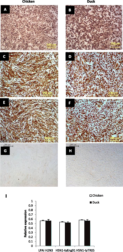
Figure 1
Figure 1
Chicken and duck cells showed comparable susceptibility to influenza virus infection and viral RNA accumulation. At 6 hpi with LPAI H2N3, H5N1 tyEng91 or H5N1 tyTR05 virus infection at MOI 1.0, similar accumulation of influenza nucleoprotein (NP) was evident in chicken (A, C, E) and duck (B, D, F) cells as detected by immunocytochemistry. Mock-infected chicken (G) and duck (H) cells show no staining. Comparable accumulation of viral matrix gene RNA between chicken and duck cells at 24 hpi at 1.0 MOI with LPAI H2N3, H5N1-tyEng91 or H5N1 tyTR05 viruses (I). Data were derived from biological replicates of 3 total RNA samples and the data points are mean relative expression values normalized to 18SrRNA expression.
Figure 2
Figure 2
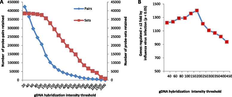
Genomic DNA (gDNA) hybridization intensity threshold of 200 provided the highest sensitivity for duck transcriptomic analysis on the chicken GeneChip. (A) The retention of whole probe-sets from duck gDNA hybridization on the chicken GeneChip array, representing transcripts, was less sensitive to the increase in gDNA hybridization intensities as only a minimum of one probe pair is required to retain a probe-set. (B) gDNA hybridization intensity threshold of 200 gave the highest number of significantly differentially regulated genes at ±2 fold (p ≤0.05) at 24 h following influenza virus infection compared with mock-infected controls. Data derived from hybridizing infected and mock-infected control duck RNA samples on chicken array.
Figure 3
Figure 3
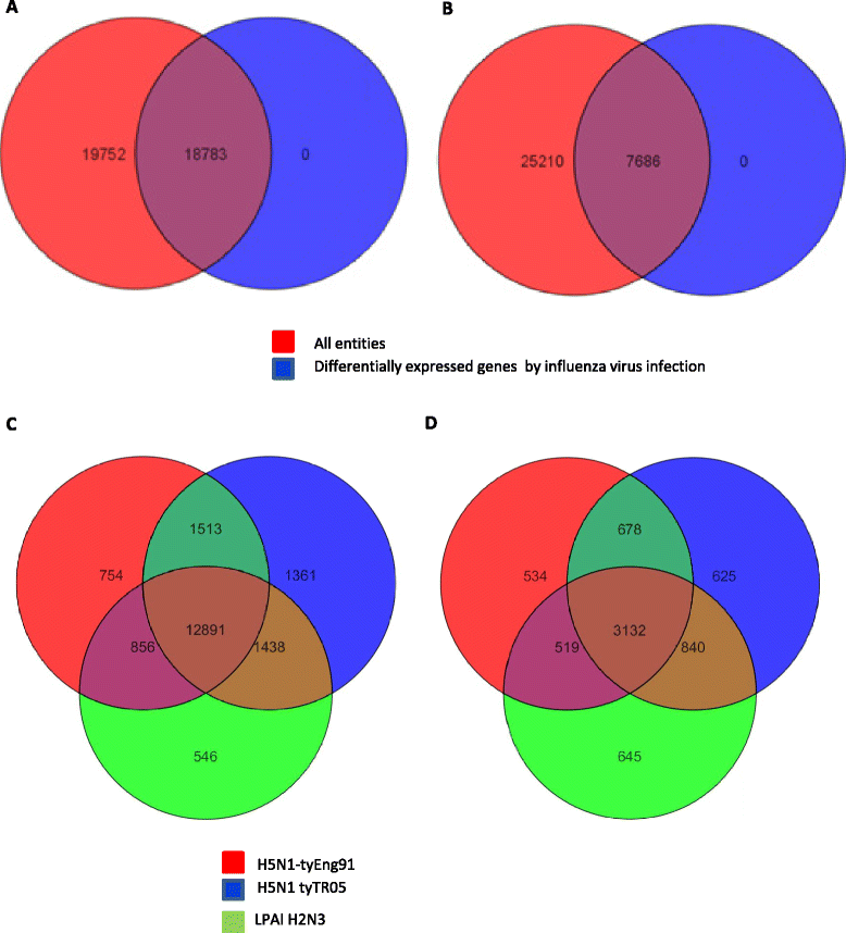
Summary of global gene expression in chicken and duck cells in response to influenza virus infection. Combined gene expression profiles of virus infected (all three avian viruses combined) and mock-infected samples showed that 18 783 out of 38 535 transcripts (48.74%) were significantly differentially regulated (P < 0.05) in chicken cells (A) while only 7686 out of 32 896 transcripts (23.36%) were significantly differentially regulated (P < 0.05) in duck cells (B) at 24 h following virus infection. Venn diagram overlap of significantly differentially regulated genes with a fold change of ±1.3 (≥ 1.3 fold, p ≤ 0.05) in (C) chicken cells and (D) duck cells at 24 h following infection with H5N1-tyEng91 (red), H5N1 tyTR05 (blue) or LPAI H2N3 (green) viruses.
Figure 4
Figure 4
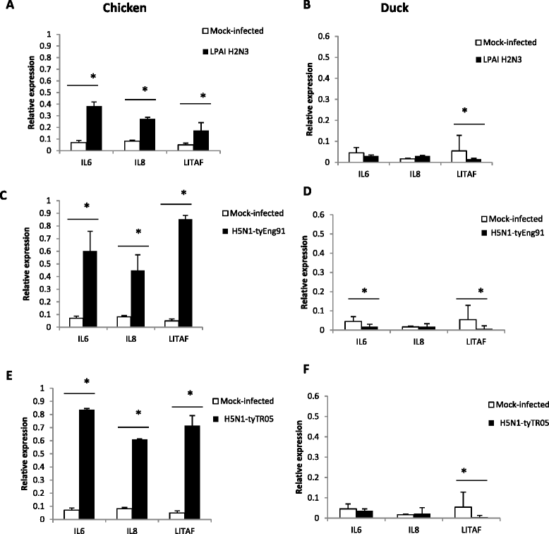
Contrasting pro-inflammatory cytokine gene response between chicken and duck cells. In chicken cells at 24 h following infection with (A) LPAI H2N3, (C) H5N1-tyEng91 or (E) H5N1-tyTR05 viruses, mRNA expression levels of IL-6, IL-8 and LITAF were significantly up-regulated. In duck cells at 24 h following infection with (B) LPAI H2N3 (D) H5N1-tyEng91 or (F) H5N1-tyTR05 viruses, IL-6, IL-8 and LITAF mRNA levels were either significantly down-regulated or unchanged. Relative mRNA expression was determined by real-time PCR normalised to 18S rRNA. Data points are the mean of three biological replicates with error bars as standard deviation (*p < 0.05).
Figure 5
Figure 5
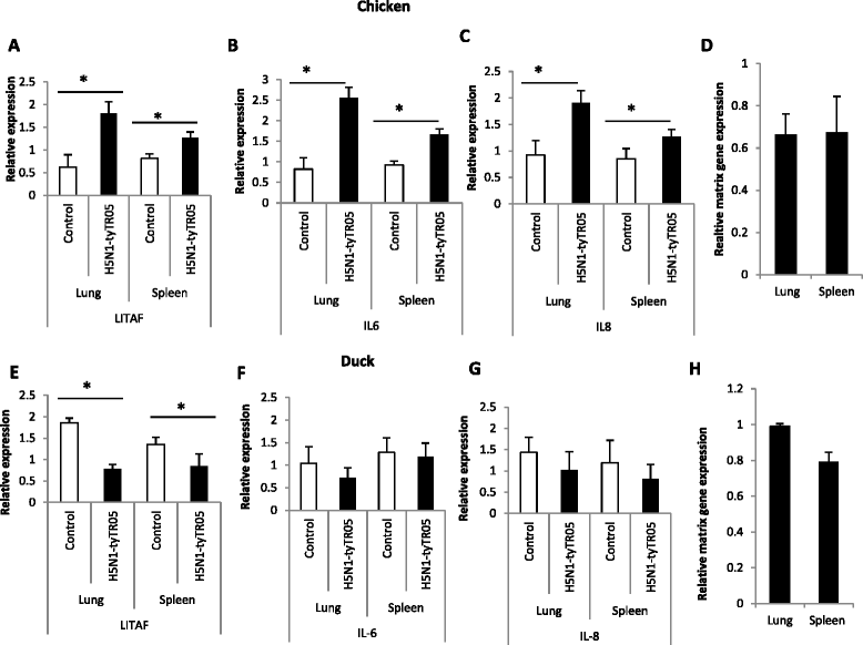
Pro-inflammatory cytokine gene response to H5N1 virus challenge in chickens and ducks. In the lungs and spleens of 3-weeks-old chickens at 24 h following infection with H5N1-tyTR05 virus, mRNA expression levels of (A) LITAF, (B) IL-6 and (C) IL-8 were significantly up-regulated compared with mock-infected controls. (D) Increased pro-inflammatory gene response in virus infected lungs correlated with RNA accumulation of influenza virus M-gene. In contrast, in the lungs and spleens of 4- weeks- old ducks infected with H5N1-tyTR05 virus, (H) despite viral M-gene RNA detection, (E) LITAF mRNA expression was significantly down-regulated and expression of (F) IL-6 and (G) IL-8 unaffected in relation to mock-infected controls. Relative mRNA expression was determined by real-time PCR normalised to 18S rRNA. Data points are the mean of three biological replicates with error bars as standard deviation.
Figure 6
Figure 6
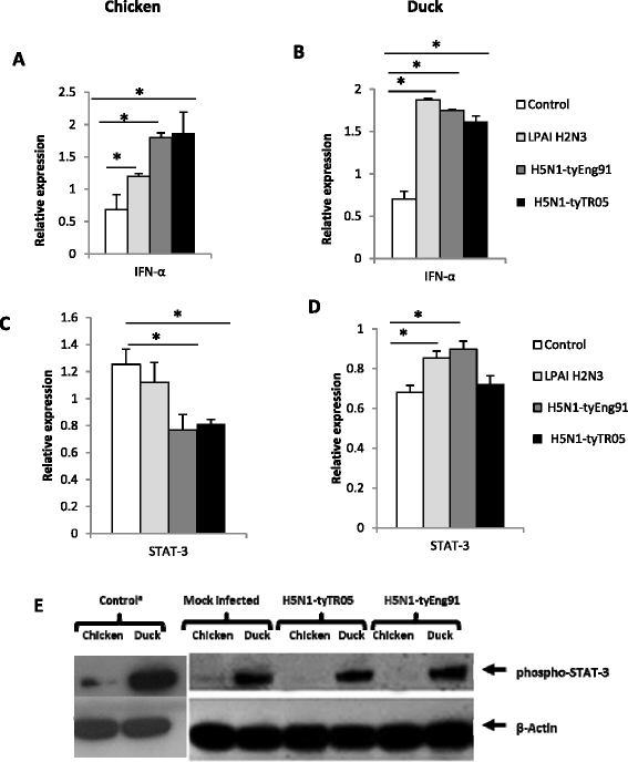
Infected chicken and duck cells showed differential regulation of STAT-3 . IFN-α expression was significantly up-regulated in chicken (A) and duck (B) cells at 24 h following infection with LPAI-H2N3, H5N1-tyEng91 or H5N1-tyTR05 viruses. (C) While STAT-3 expression in chicken cells was not significantly affected by LPAI-H2N3 virus infection it was significantly down-regulated in H5N1-tyEng91 and H5N1-tyTR05 virus infections. (D) In contrast STAT-3 expression in duck cells was significantly up-regulated by LPAI-H2N3 or H5N1-tyEng91 viruses but was not affected by H5N1-tyTR05 virus infection. Relative mRNA expression was determined by real-time PCR normalised to 18S rRNA. Data points are the mean of three biological replicates with error bars as standard deviation (*p < 0.05). (E) Strong constitutive phospho-STAT-3 protein expression was detected in duck cells which was unaffected at 24 h following infection with H5N1-tyEng91 and H5N1-tyTR05 viruses. In chicken mock-infected cells, phospho-STAT3 protein expression was scarcely detectable and remained absent at 24 h following virus infection. aLonger (5 min) exposure showing pSTAT-3 in chicken cells.
Figure 7
Figure 7
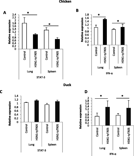
Differential STAT-3 regulation between H5N1 virus-infected chickens and ducks. In the lungs and spleens of 3-weeks-old chickens at 24 h following infection with H5N1-tyTR05 viruses, (A) expression of STAT-3 was significantly down-regulated whereas (B) IFN-α was significantly up-regulated. In the lung and spleen tissues from 4- weeks- old ducks at 24 h following infection with H5N1-tyTR05 virus, (C) STAT-3 expression was unaffected and (D) IFN-α expression was significantly up-regulated. Relative mRNA expression was determined by real-time PCR normalised to 18S rRNA. Data points are the mean of three biological replicates with error bars as standard deviation (*p < 0.05).
Figure 8
Figure 8
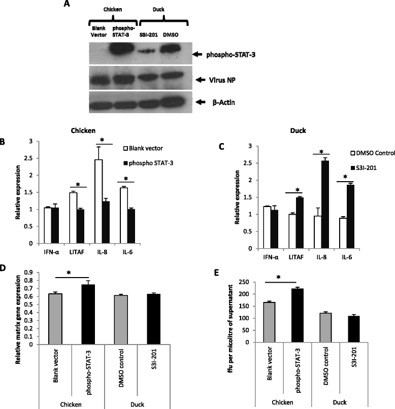
STAT-3 appears to regulate the pro-inflammatory response and promote virus replication in H5N1 virus infected chicken and duck cells. (A) Primary chicken embryo cells over-expressing phospho-STAT-3 showed a high phospho-STAT-3 expression while STAT-3 inhibitor S3I-201 treatment resulted in reduced phospho-STAT-3 protein expression in duck cells at 24 h following H5N1-tyEng91 virus infection (1.0 MOI). (B) phospho-STAT-3 over-expressing chicken cells showed a significant reduction in LITAF, IL6 and IL-8 mRNA expression with no significant change in IFN-α expression. (C) At 24 h following H5N1-tyEng91 virus infection, in STAT-3 inhibited duck primary embryo cells, significant increase of LITAF, IL-8 and IL-6 mRNA expression was detected with no significant change in IFN-α expression. Phospho STAT-3 over-expression in chicken cells increased viral replication at 24 h following H5N1-tyEng91 virus infection as evidenced by increased detection of virus NP (A), matrix gene mRNA (D) and infectious virus output in culture supernatant (E). STAT-3 inhibition did not significantly affect virus NP (A) matrix gene expression (D) or infectious virus production (E) at 24 h following H5N1-tyEng91 virus infection in duck cells. Relative mRNA expression was determined by real-time PCR to 18S rRNA. Data points are the mean of three biological replicates with error bars as standard deviation (*p < 0.05).

Similar articles

Cited by

References

    1. Gibbs AJ, Armstrong JS, Downie JC. From where did the 2009 ‘swine-origin’ influenza A virus (H1N1) emerge? Virol J. 2009;6:207. doi: 10.1186/1743-422X-6-207. - DOI - PMC - PubMed
    1. Thitithanyanont A, Engering A, Uiprasertkul M, Ekchariyawat P, Wiboon-Ut S, Kraivong R, Limsalakpetch A, Kum-Arb U, Yongvanitchit K, Sa-Ard-Iam N, Rukyen P, Mahanonda R, Kawkitinarong K, Auewarakul P, Utaisincharoen P, Sirisinha S, Mason CJ, Fukuda MM, Pichyangkul S. Antiviral immune responses in H5N1-infected human lung tissue and possible mechanisms underlying the hyperproduction of interferon-inducible protein IP-10. Biochem Biophys Res Commun. 2010;398:752–758. doi: 10.1016/j.bbrc.2010.07.017. - DOI - PMC - PubMed
    1. Katz JM, Veguilla V, Belser JA, Maines TR, Van Hoeven N, Pappas C, Hancock K, Tumpey TM. The public health impact of avian influenza viruses. Poult Sci. 2009;88:872–879. doi: 10.3382/ps.2008-00465. - DOI - PubMed
    1. Sharp GB, Kawaoka Y, Jones DJ, Bean WJ, Pryor SP, Hinshaw V, Webster RG. Coinfection of wild ducks by influenza A viruses: distribution patterns and biological significance. J Virol. 1997;71:6128–6135. - PMC - PubMed
    1. Isoda N, Sakoda Y, Kishida N, Bai GR, Matsuda K, Umemura T, Kida H. Pathogenicity of a highly pathogenic avian influenza virus, A/chicken/Yamaguchi/7/04 (H5N1) in different species of birds and mammals. Arch Virol. 2006;151:1267–1279. doi: 10.1007/s00705-005-0723-6. - DOI - PubMed

Publication types

MeSH terms

Associated data