. 2014 Sep;9(9):863-876.
doi: 10.2217/fvl.14.66.

The RNA-dependent RNA polymerase of the influenza A virus

Affiliations

The RNA-dependent RNA polymerase of the influenza A virus

Thomas M Stubbs et al. Future Virol. 2014 Sep.

Abstract

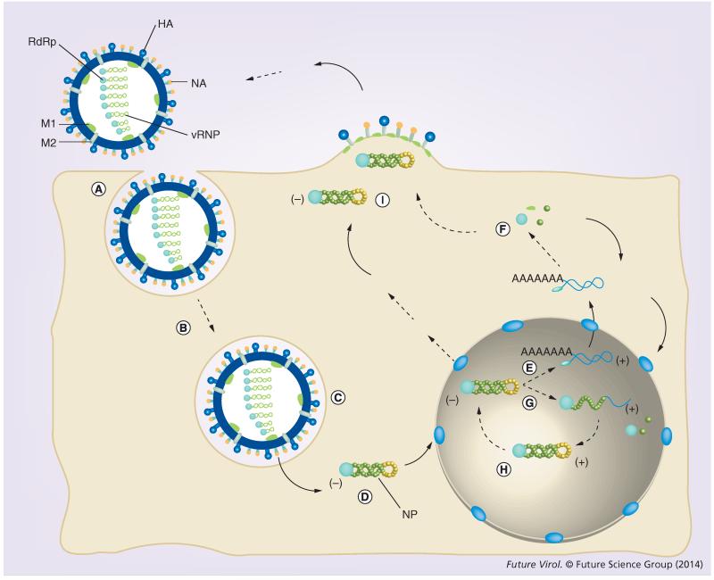
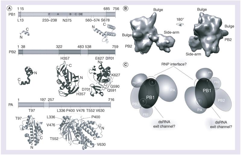
The influenza A virus causes a highly contagious respiratory disease that significantly impacts our economy and health. Its replication and transcription is catalyzed by the viral RNA polymerase. This enzyme is also crucial for the virus, because it is involved in the adaptation of zoonotic strains. It is thus of major interest for the development of antiviral therapies and is being intensively studied. In this article, we will discuss recent advances that have improved our knowledge of the structure of the RNA polymerase and how mutations in the polymerase help the virus to spread effectively among new hosts.

Keywords: RNA polymerase; adaptation; host restriction; influenza A; replication; structure; transcription.


Figures

Figure 1
Figure 1. Influenza A virus replication cycle
(A) A typical IAV infection starts with the binding of the HA proteins on the surface of the virus particle to the sialic acid-containing oligosaccharides on the surface of the host cell. Human influenza strains typically bind α2,6-linked sialic acid, which is present in the upper respiratory tract, while avian strains recognize α2,3-linked sialic acid, which is present in the lower respiratory tract [97]. (B) Binding to the host receptors is followed by endocytosis, (C) fusion of the endosomal membranes with the membrane of the virion and release of the vRNPs. (D) vRNPs are next transported to the nucleus of the host. NLSs on the viral proteins and host importins play a crucial role in this process. (E) In the nucleus, transcription is initiated with capped primers that are cleaved from cellular pre-mRNAs. (F) Mature viral mRNAs are exported to the cytoplasm for the synthesis of new viral proteins. (G) New viral RNP subunits are imported into the nucleus and associate with newly forming cRNAs in order to create cRNPs. (H) cRNPs next synthesize new vRNAs. (I) After several rounds of amplification, newly formed vRNPs are exported from the nucleus and transported to the cell membrane in a Rab11- and microtubule-dependent manner for packaging and budding. cRNA: Complementary RNA; cRNP: Complementary ribonucleoprotein; HA: Hemagglutinin; IAV: Influenza A virus; NLS: Nuclear localization signal; vRNA: Viral RNA; vRNP: Viral ribonucleoprotein.
Figure 2
Figure 2. Structure of the influenza A virus RNA-dependent RNA polymerase
(A) The influenza A virus (IAV) RNA-dependent RNA polymerase (RdRp) consists of the subunits PA, PB1 and PB2. Six of the seven canonical RdRp domains motifs A–F are found in PB1. Also indicated are the RNA binding sequences (gray), as are the residues involved in adaptation (black; see also Table 1). At present, significant structural information is only available for PA and PB2, which is shown below the schematics of the subunits. The visualized structural information is based on Protein Data Bank (PDB) entries 2VY6, 2W69, 2ZNL, 2ZTT, 3EBJ and 4CB4. (B) Two views of the electron microscopy model proposed by Moeller et al. [39]. Panels are based on the PDBe entry EMD-2213. (C) Two interpretations based on current electron microscopy models. On the left, the PA C-terminus presents the flexible part of the IAV RdRp, as defined by Moeller et al. [39]. It is connected to the PA endonuclease domain via a linker [37], which may wrap around PB1. An interaction through the core of PB1 would seem unlikely, as this may interfere with the right-handed fold of the polymerase active site. On the right, the PB2 C-terminus presents the flexible part of the IAV RdRp, as proposed by Torreira et al. [49], The PA domains flank PB1 on the other side, but the PA N-terminus can still interact with PB2 in accordance with recent data [47]. In both models, the flexible domain can be envisioned as blocking the dsRNA exit channel of the RdRp, which likely resides opposite – albeit at a slight angle – to the template entry grove. Both interpretations also have a similar interface consisting of PB1 and PA residues in order to interact with the RNP. Since the PA linker domain may not be part of this interaction, it is displayed in both models as a dashed line. The areas in (C) labeled with question marks refer to the bulge structures in (B), whose identity is current unknown. PA: Polymerase acidic; PB1: Polymerase basic 1; PB2: Polymerase basic 2; RNP: Ribonucleoprotein.

References

    1. Taubenberger JK, Morens DM. Pandemic influenza – including a risk assessment of H5N1. Rev. Sci. Tech. 2009;28(1):187–202. - PMC - PubMed
    1. Presti RM, Zhao G, Beatty WL, et al. Quaranfil, Johnston Atoll, and Lake Chad viruses are novel members of the family Orthomyxoviridae. J. Virol. 2009;83(22):11599–11606. - PMC - PubMed
    1. Girard MP, Tam JS, Assossou OM, Kieny MP. The 2009 A (H1N1) influenza virus pandemic: a review. Vaccine. 2010;28(31):4895–4902. - PubMed
    1. Das K. Antivirals targeting influenza A virus. J. Med. Chem. 2012;55(14):6263–6277. - PubMed
    1. Ekiert DC, Wilson IA. Broadly neutralizing antibodies against influenza virus and prospects for universal therapies. Curr. Opin. Virol. 2012;2(2):134–141. - PMC - PubMed

LinkOut - more resources