

Variations in eyeball diameters of the healthy adults
- PMID: 25431659
- PMCID: PMC4238270
- DOI: 10.1155/2014/503645
Variations in eyeball diameters of the healthy adults
Abstract
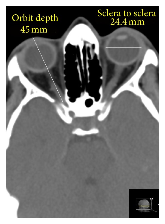
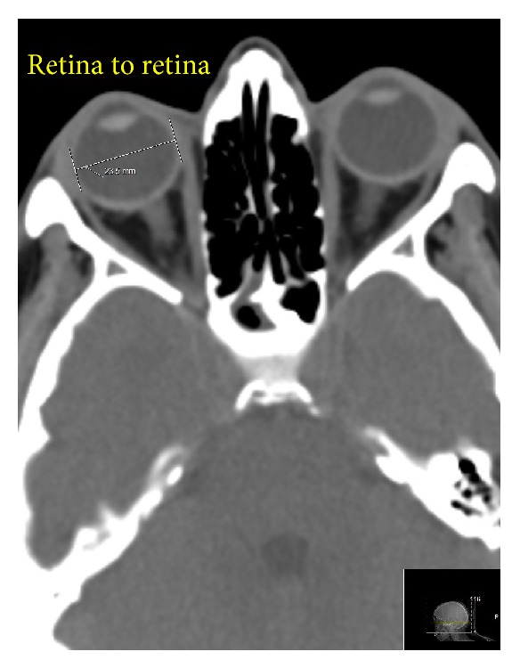
The purpose of the current research was to reevaluate the normative data on the eyeball diameters. Methods. In a prospective cohort study, the CT data of consecutive 250 adults with healthy eyes were collected and analyzed, and sagittal, transverse, and axial diameters of both eyeballs were measured. The data obtained from the left eye and from the right eye were compared. The correlation analysis was performed with the following variables: orbit size, gender, age, and ethnic background. Results. We did not find statistically significant differences correlated with gender of the patients and their age. The right eyeball was slightly smaller than the left one but this difference was statistically insignificant (P = 0.17). We did not find statistically significant differences of the eyeball sizes among the ethnicities we dealt with. Strong correlation was found between the transverse diameter and the width of the orbit (r = 0.88). Conclusion. The size of a human adult eye is approximately 24.2 mm (transverse) × 23.7 mm (sagittal) × 22.0-24.8 mm (axial) with no significant difference between sexes and age groups. In the transverse diameter, the eyeball size may vary from 21 mm to 27 mm. These data might be useful in ophthalmological, oculoplastic, and neurological practice.
Figures
References
-
- Kim J. M., Kim M. S., Jang H. J., Park K. H., Caprioli J. The association between retinal vessel diameter and retinal nerve fiber layer thickness in asymmetric normal tension glaucoma patients. Investigative Ophthalmology and Visual Science. 2012;53(9):5609–5614. doi: 10.1167/iovs.12-9783. - DOI - PubMed
-
- Liebovitch L. S. Why the eye is round? In: Fischbarg J., editor. The Biology of the Eye. Boston, Mass, USA: Elsevier; 2006. pp. 5–6.
LinkOut - more resources
Full Text Sources
Other Literature Sources
