Spontaneous intraperitoneal rupture of a hepatic hydatid cyst with subsequent anaphylaxis: a case report
- PMID: 25431702
- PMCID: PMC4238233
- DOI: 10.1155/2013/320418
Spontaneous intraperitoneal rupture of a hepatic hydatid cyst with subsequent anaphylaxis: a case report
Abstract
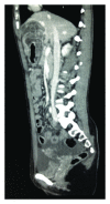
Hydatid cyst rupture into the abdomen is a serious complication of cystic hydatid disease of the liver (Cystic Echinococcosis) with an incidence of up to 16% in some series and can result in anaphylaxis or anaphylactoid reactions in up to 12.5% of cases. At presentation, 36-40% of hydatid cysts have ruptured or become secondarily infected. Rupture can be microscopic or macroscopic and can be fatal without surgery. Hydatid disease of the liver is primarily caused by the tapeworm Echinococcus granulosus and occurs worldwide, with incidence of up to 200 per 100,000 in endemic areas. Our case describes a 24-year-old Bulgarian woman presenting with epigastric pain and evidence of anaphylaxis. Abdominal CT demonstrated a ruptured hydatid cyst in the left lobe of the liver. A partial left lobe hepatectomy, cholecystectomy, and peritoneal washout was performed with good effect. She was treated for anaphylaxis and received antihelminthic treatment with Albendazole and Praziquantel. She made a good recovery following surgery and medical treatment and was well on follow-up. Intraperitoneal rupture with anaphylaxis is a rare occurrence, and there do not seem to be any reported cases from UK centres prior to this.
Figures


References
-
- McIntyre N, Benhamou J-P, Bircher J, Rizzetto M, Rodes J, editors. Oxford Textbook of Clinical Hepatology. 1991;1
-
- Sherlock S, Dooley J. Diseases of the Liver and Biliary System. 11th edition 2002.
-
- Moro P, Schantz PM. Echinococcosis: a review. International Journal of Infectious Diseases. 2009;13(2):125–133. - PubMed
-
- Akcan A, Akyildiz H, Artis T, et al. Peritoneal perforation of liver hydatid cysts: clinical presentation, predisposing factors, and surgical outcome. World Journal of Surgery. 2007;31(6):1284–1291. - PubMed
LinkOut - more resources
Full Text Sources
Other Literature Sources

