. 2015 Feb 15;466(1):137-45.
doi: 10.1042/BJ20141237.

A directed-overflow and damage-control N-glycosidase in riboflavin biosynthesis

Affiliations

A directed-overflow and damage-control N-glycosidase in riboflavin biosynthesis

Océane Frelin et al. Biochem J. .

Abstract

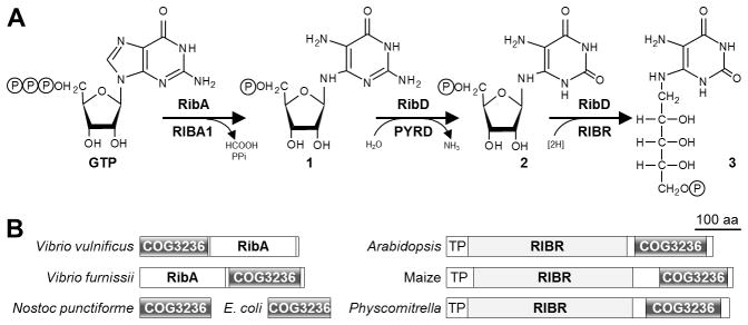
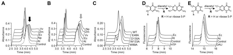
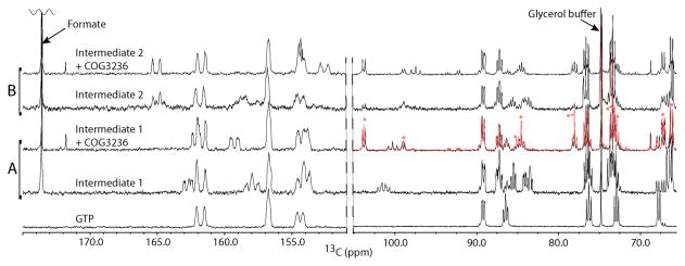
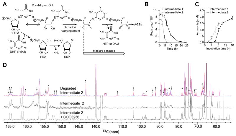
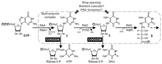
Plants and bacteria synthesize the essential human micronutrient riboflavin (vitamin B2) via the same multi-step pathway. The early intermediates of this pathway are notoriously reactive and may be overproduced in vivo because riboflavin biosynthesis enzymes lack feedback controls. In the present paper, we demonstrate disposal of riboflavin intermediates by COG3236 (DUF1768), a protein of previously unknown function that is fused to two different riboflavin pathway enzymes in plants and bacteria (RIBR and RibA respectively). We present cheminformatic, biochemical, genetic and genomic evidence to show that: (i) plant and bacterial COG3236 proteins cleave the N-glycosidic bond of the first two intermediates of riboflavin biosynthesis, yielding relatively innocuous products; (ii) certain COG3236 proteins are in a multi-enzyme riboflavin biosynthesis complex that gives them privileged access to riboflavin intermediates; and (iii) COG3236 action in Arabidopsis thaliana and Escherichia coli helps maintain flavin levels. COG3236 proteins thus illustrate two emerging principles in chemical biology: directed overflow metabolism, in which excess flux is diverted out of a pathway, and the pre-emption of damage from reactive metabolites.


Figures

Figure 1
Figure 1. Evidence connecting COG3236 with riboflavin biosynthesis
(A) The first three steps of riboflavin biosynthesis and their enzymes in bacteria (above arrows) and plants (below arrows). Steps two and three in bacteria are catalyzed by RibD, a bifunctional deamin-ase-reductase. RibA and RIBA1, GTP cyclohydrolase II; RibD and PYRD, pyrimidine deaminase; RibD and RIBR, pyrimidine reductase. Intermediates 1, 2, and 3 are 2,5-diamino-6-ribosylamino-4(3H)-pyrimidinone 5′-phosphate, 5-amino-6-ribosylamino-2,4(1H,3H)-pyrimidinedione 5′-phosphate, and 5-amino-6-ribitylamino-2,4(1H,3H)-pyrimidinedione 5′-phosphate, respectively; P, phosphate. (B) Domain structures of fusions between COG3236 and RibA in γ-Proteobacteria, and between COG3236 and RIBR in plants. Two stand-alone bacterial COG3236 proteins are also shown. TP, plastid targeting peptide.
Figure 2
Figure 2. Chromatographic evidence that plant and bacterial COG3236 proteins cleave the pyrimidine moiety from riboflavin biosynthesis intermediates 1 and 2
(A) Action of E. coli (Ec), N. punctiforme (Np), V. vulnificus (Vv), Arabidopsis (At), and maize (Zm) COG3236 proteins on intermediate 1 (black arrow). Reactions (50 μl) containing 3 mM intermediate 1 and 30 μg of protein were incubated for 1 h at 22°C and analyzed by HPLC with UV detection. Control reactions contained no enzyme. (B) Action of COG3236 proteins on intermediate 2 (grey arrow). Conditions were as in A except that substrate concentration was 2.4 mM and the amounts of protein (μg) were: Vv, 5; Ec and Np, 1; Zm 0.25; At, 0.1. The shoulder on the intermediate 2 peak is due to partial resolution of the two anomers [9]. (C) Elimination or reduction of activity of E. coli COG3236 by site-directed mutagenesis of four conserved residues. A reaction with wild type (WT) protein is shown for comparison. Conditions were as in A except that 100 μg of protein was used. (D,E) Fluorometric HPLC analysis of the diacetyl derivatives of, respectively, intermediates 1 and 2, of the products formed therefrom by E. coli or Arabidopsis COG3236, and of authentic 4-hydroxy-2,5,6-triaminopyrimidine (HTP) or 5,6-diaminouracil (DAU). Control reactions contained no enzyme.
Figure 3
Figure 3. NMR analysis confirms that E. coli COG3236 cleaves the N-glycosidic bond of riboflavin intermediates 1 and 2
Aromatic (left) and ribose regions (right) are shown for reactions involving (A) intermediate 1 generated by RibA and (B) intermediate 2 generated by RibA plus RibD. The starting GTP is shown as reference at the bottom. Spectra were obtained using volumes of just 50 μl by incubating labeled GTP with COG3236 in the 1.5-mm NMR tubes. An exceptionally sensitive custom 13C-optimized NMR probe made from high temperature superconducting material [56] was utilized. The red overlay in the ribose region of ‘Intermediate 1 + COG3236’ is the ribose-5-phosphate spike of the same spectrum in black. Asterisks indicate the ribose-5-phosphate resonances that increased in the spike. Note that ribose-5-phosphate exists as a mixture of α and β anomers. Assignments of NMR spectra of unstable intermediates were made with calculated chemical shifts (Table S1).
Figure 4
Figure 4. Spontaneous breakdown chemistry of intermediates 1 and 2
(A) Hypothetical breakdown reactions of intermediates 1 and 2. 5AB, 5-aminobarbituric acid; AGEs, advanced glycation end products; DAU, diaminouracil; DHP, 2,5-diamino-4,6-dihydroxypyrimidine; HTP, 4-hydroxy-2,5,6-triaminopyrimidine; PRA, 5-phosphoribosylamine; R-5-P, ribose-5-phosphate. (B) Kinetics of the disappearance of the UV-absorbing HPLC peaks of intermediates 1 and 2. (C) Kinetics of NH3 release from intermediates 1 (3 mM) and 2 (2.4 mM). (D) Freshly prepared 13C,15N-labeled intermediate 2 (2.4 mM) was incubated at 22°C in the NMR tube and 13C NMR spectra were acquired during successive 4-h periods. The first spectrum (blue) and last spectrum collected at 20 h (red) are shown at the top. The spectra from Figure 3 of intermediate 2 (middle) and intermediate 2 + COG3236 (bottom) are shown for comparison. The left portion is the aromatic region and the right portion is the ribose region. The arrows above the degradation spectra indicate resonances that decrease (↓) or increase (↑) over time. Spectral changes for intermediate 1 were similar but less marked.
Figure 5
Figure 5. A model of COG3236 action in the riboflavin biosynthesis pathway
COG3236 proteins dispose of excess intermediates 1 and 2 by hydrolysis to ribose-5-phosphate and a pyrimidine. These are less damaging than compounds such as Maillard products and 5-phospho-ribosylamine (PRA) to which the intermediates could spontaneously break down. The pathway enzymes, and COG3236, form a multi-enzyme complex. HTP, 4-hydroxy-2,5,6-triaminopyrimidine; DAU, 5,6-diaminouracil.

References

    1. Reaves ML, Young BD, Hosios AM, Xu YF, Rabinowitz JD. Pyrimidine homeostasis is accomplished by directed overflow metabolism. Nature. 2013;500:237–241. - PMC - PubMed
    1. Linster CL, Van Schaftingen E, Hanson AD. Metabolite damage and its repair or pre-emption. Nat Chem Biol. 2013;9:72–80. - PubMed
    1. Vinci CR, Clarke SG. Homocysteine methyltransferases Mht1 and Sam4 prevent the accumulation of age-damaged (R,S)-AdoMet in the yeast Saccharomyces cerevisiae. J Biol Chem. 2010;285:20526–20531. - PMC - PubMed
    1. Danchin A, Sekowska A. The logic of metabolism and its fuzzy consequences. Environ Microbiol. 2014;16:19–28. - PubMed
    1. Golubev AG. The other side of metabolism: a review. Biochemistry (Mosc) 1996;61:2018–2039. - PubMed

Publication types

MeSH terms