. 2015 Jun;290(3):847-62.
doi: 10.1007/s00438-014-0959-5. Epub 2014 Nov 29.

RNase E in the γ-Proteobacteria: conservation of intrinsically disordered noncatalytic region and molecular evolution of microdomains

Affiliations

RNase E in the γ-Proteobacteria: conservation of intrinsically disordered noncatalytic region and molecular evolution of microdomains

Soraya Aït-Bara et al. Mol Genet Genomics. 2015 Jun.

Abstract

RNase E of Escherichia coli is a membrane-associated endoribonuclease that has a major role in mRNA degradation. The enzyme has a large C-terminal noncatalytic region that is mostly intrinsically disordered (ID). Under standard growth conditions, RhlB, enolase and PNPase associate with the noncatalytic region to form the multienzyme RNA degradosome. To elucidate the origin and evolution of the RNA degradosome, we have identified and characterized orthologs of RNase E in the γ-Proteobacteria, a phylum of bacteria with diverse ecological niches and metabolic phenotypes and an ancient origin contemporary with the radiation of animals, plants and fungi. Intrinsic disorder, composition bias and tandem sequence repeats are conserved features of the noncatalytic region. Composition bias is bipartite with a catalytic domain proximal ANR-rich region and distal AEPV-rich region. Embedded in the noncatalytic region are microdomains (also known as MoRFs, MoREs or SLiMs), which are motifs that interact with protein and other ligands. Our results suggest that tandem repeat sequences are the progenitors of microdomains. We have identified 24 microdomains with phylogenetic signals that were acquired once with few losses. Microdomains involved in membrane association and RNA binding are universally conserved suggesting that they were present in ancestral RNase E. The RNA degradosome of E. coli arose in two steps with RhlB and PNPase acquisition early in a major subtree of the γ-Proteobacteria and enolase acquisition later. We propose a mechanism of microdomain acquisition and evolution and discuss implications of these results for the structure and function of the multienzyme RNA degradosome.


Figures
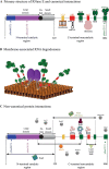
Fig. 1
Fig. 1
E. coli RNase E is a protein interaction hub. a Primary structure of E. coli RNase E showing the catalytic region (residues 1–529) and the noncatalytic region (residues 530–1,061 residues). The catalytic region is composed of a large domain, a zinc-link (Zn-link) and a small domain. The large domain is contains an S1 RNA binding motif (blue Pfam00575) and a metal-binding catalytic site (purple, Pfam10150). The RNase E tetrameric holoenzyme is a dimer of dimers. The large domain and Zn-link have a structural role in dimer formation; the small domain has a structural role in dimer and dimer–dimer interactions. The noncatalytic region (residues 530–1,061) contains microdomains responsible for the interaction between the RNase E and the inner plasma membrane (yellow MTS, membrane targeting sequence, residues 565–582), RNA (red AR1, arginine-rich 1, residues 604–644; AR2, arginine-rich 2, residues 796–814), and proteins to form the canonical RNA degradosome (green HBS, helicase binding site, residues 719–731; EBS, enolase binding site, residues 834–850 residues; PBS (Pfam12111), PNPase binding site, residues 1,021–1,061 residues). b Cartoon showing tetrameric RNase E holoenzyme bound to the inner cytoplasmic membrane and organization of the RNA degradosome. Purple catalytic core of RNase E; gray ID region; yellow MTS; red RNA binding sites; green protein binding sites and associated proteins. c Non-canonical protein interactions with E. coli RNase E. Hfq (residues 711–750) (Ikeda et al. 2011); CsdA, SrmB and RhlE (residues 791–843) (Khemici et al. ; Prud’homme-Genereux et al. 2004); RraA (residues 604–688 and 791–819) and RraB (residues 694–727) (Gao et al. ; Gorna et al. 2010); MinD (residues 378–724) (Taghbalout and Rothfield 2007); RapZ (residues 1–529) (Gopel et al. 2013); poly(A)polymerase (PAPI) (residues 501–843) (Raynal and Carpousis ; Carabetta et al. 2010). Other non-canonical interactions have been mentioned in the literature but the binding sites are unknown: RNase R (Carabetta et al. 2010); GroEL, DnaK, and polyphosphate kinase (PPK) (Miczak et al. ; Blum et al. 1997); ribosomal proteins such as S1, L4, L17 (Feng et al. ; Singh et al. ; Tsai et al. 2012) (color figure online)
Fig. 2
Fig. 2
RNase E and RNase G form distinct orthologous groups in the γ-Proteobacteria. Phylogenetic trees of RNase E and RNase G homologs were constructed as described (“Materials and methods”). Gray dots indicate branches with high bootstrap support. Inner circle tree leaves colored according to the taxonomy (taxonomy key). Center circle RNase G, red; RNase E, purple. Outer circle line diagram of primary structure of RNase E and RNase G homologs showing the conserved Pfam domains (protein key) (color figure online)
Fig. 3
Fig. 3
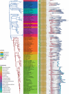
Distribution of RNase G, RNase E, PNPase, enolase and RNase R. The phylogenetic tree of γ-Proteobacteria species was constructed as described (“Materials and methods”). The blue branches correspond to a subdivision that includes the PO clade (Pseudomonadales and Oceanospirillales); the red branches to the VAAP clade (Vibrionales, Aeromonadales, Alteromonadales, Pasteurellales) and Enterobacteriales. Inner circle tree leaves according to the taxonomy (taxonomy key). Center circle red dots indicate genome size. Outer circle distribution of proteins (protein key). The gene encoding enolase is present in multiple copies in Marinobacter adhaerens and Azotobacter vinelandii. A second copy of the gene encoding RNase R is present in a few species and the protein is labeled RNase Rb (color figure online)
Fig. 4
Fig. 4
Composition bias in the noncatalytic region of RNase E orthologs. Primary structure of a representative selection of RNase E homologs (right half of panel) is mapped to the species tree of the γ-Proteobacteria (left half of panel), which was constructed as described (“Materials and methods”). The blue branches correspond to a subtree that includes the PO clade (Pseudomonadales and Oceanospirillales); the red branches to the VAAP clade (Vibrionales, Aeromonadales, Alteromonadales, Pasteurellales) and Enterobacteriales. Tree leaves are color coded according to taxonomy (key). Symbols for Pfam domains and composition bias (CB) are indicated in the protein key. Note that the symbols for CB represent the region of bias, they do not imply a gradient or directionality (color figure online)
Fig. 5
Fig. 5
Conserved sequence motifs in the noncatalytic region of RNase E orthologs. The primary structure of a representative selection of RNase E homologs (right half of panel) is mapped to the species tree of the γ-Proteobacteria (left half of panel), which was constructed as described (“Materials and methods”). The blue branches correspond to a subdivision that includes the PO clade (Pseudomonadales and Oceanospirillales); the red branches to the VAAP clade (Vibrionales, Aeromonadales, Alteromonadales, Pasteurellales) and Enterobacteriales. Tree leaves are color coded according to taxonomy (key). Symbols for Pfam domains and conserved sequence motifs are indicated in the protein key (color figure online)

References

    1. Abascal F, Zardoya R, Posada D. ProtTest: selection of best-fit models of protein evolution. Bioinformatics. 2005;21:2104–2105. doi: 10.1093/bioinformatics/bti263. - DOI - PubMed
    1. Ait-Bara S, Carpousis AJ. Characterization of the RNA degradosome of Pseudoalteromonas haloplanktis: conservation of the RNase E-RhlB interaction in the {gamma}-Proteobacteria. J Bacteriol. 2010;192:5413–5423. doi: 10.1128/JB.00592-10. - DOI - PMC - PubMed
    1. Alba MM, Laskowski RA, Hancock JM. Detecting cryptically simple protein sequences using the SIMPLE algorithm. Bioinformatics. 2002;18:672–678. doi: 10.1093/bioinformatics/18.5.672. - DOI - PubMed
    1. Altschul SF, Madden TL, Schaffer AA, Zhang J, Zhang Z, Miller W, Lipman DJ. Gapped BLAST and PSI-BLAST: a new generation of protein database search programs. Nucleic Acids Res. 1997;25:3389–3402. doi: 10.1093/nar/25.17.3389. - DOI - PMC - PubMed
    1. Bailey TL, Williams N, Misleh C, Li WW. MEME: discovering and analyzing DNA and protein sequence motifs. Nucleic Acids Res. 2006;34:W369–W373. doi: 10.1093/nar/gkl198. - DOI - PMC - PubMed

Publication types

LinkOut - more resources