. 2014 Nov 27;6(12):3238-51.
doi: 10.1093/gbe/evu257.

Bioenergetic evolution in proteobacteria and mitochondria

Affiliations

Bioenergetic evolution in proteobacteria and mitochondria

Mauro Degli Esposti. Genome Biol Evol. .

Abstract

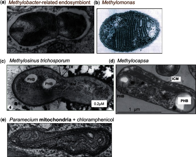
Mitochondria are the energy-producing organelles of our cells and derive from bacterial ancestors that became endosymbionts of microorganisms from a different lineage, together with which they formed eukaryotic cells. For a long time it has remained unclear from which bacteria mitochondria actually evolved, even if these organisms in all likelihood originated from the α lineage of proteobacteria. A recent article (Degli Esposti M, et al. 2014. Evolution of mitochondria reconstructed from the energy metabolism of living bacteria. PLoS One 9:e96566) has presented novel evidence indicating that methylotrophic bacteria could be among the closest living relatives of mitochondrial ancestors. Methylotrophs are ubiquitous bacteria that live on single carbon sources such as methanol and methane; in the latter case they are called methanotrophs. In this review, I examine their possible ancestry to mitochondria within a survey of the common features that can be found in the central and terminal bioenergetic systems of proteobacteria and mitochondria. I also discuss previously overlooked information on methanotrophic bacteria, in particular their intracytoplasmic membranes resembling mitochondrial cristae and their capacity of establishing endosymbiotic relationships with invertebrate animals and archaic plants. This information appears to sustain the new idea that mitochondrial ancestors could be related to extant methanotrophic proteobacteria, a possibility that the genomes of methanotrophic endosymbionts will hopefully clarify.

Keywords: bioenergetics; endosymbiosis; methanotrophs; methylotrophs; mitochondria.


Figures

F<sc>ig</sc>. 1.—
Fig. 1.—
Classification and distribution of COX operons in proteobacteria. (A) COX operons defined by Degli Esposti et al. (2014) are graphically represented with the core genes COX1–4 (corresponding to ctaBCDE in Bacillus) and common other genes including those for assembling the catalytic groups of the oxidase: ctaA (star with A), ctaB (pentagon with B), ctaG (hexagon with G), and SURF-1 (grey square). The sequences falling within COX operon type a can be distinguished from the position of ctaG (supplementary fig. S1, Supplementary Material online). The canonical gene sequence CyoA–E of bo3-type ubiquinol oxidases is shown for comparison. See Matsutani et al. (2014) for a classification of these sequences, which are clearly different from those of COX operons (see also supplementary figs. S1 and S2, Supplementary Material online). (B) Distribution of COX operon type a in the classes of α-, β-, and γ-proteobacteria. Data were extracted from NCBI websites, accessed on September 15, 2014. The total number of species with available genomic data is comparable for α- and γ-proteobacteria (589 and 652, respectively), but approximately one-half (301) for β-proteobacteria. Act, alternative complex three (Refojo et al. 2010); PQQ, deH, methanol dehydrogenase.
F<sc>ig</sc>. 2.—
Fig. 2.—
Phylogenetic tree of COX3 and evolutionary path of COX operons. The sequence of Tistrella COX3 (accession: YP_006372231, 269 residues long) was used as the query of a DELTABLAST search expanded to 5,000 proteins, including β- and γ-proteobacterial taxa for a total selection of 70 species. The Methylomicrobium group of five γ-proteobacterial taxa that is expanded on the right is the same in the branch of COX operon a–b transition and the subbranch of COX-operon a-II (arrow), which appears to be the immediate ancestral group to that containing mitochondria (enclosed in big square—see also supplementary fig. S2, Supplementary Material online). Only Glaceicola contains the same type of protein among other γ-proteobacteria, but in a COX operon that does not show methanol or related dehydrogenases (supplementary fig. S2, Supplementary Material online). The α-proteobacterial taxa in the same group include Devosia sp. DBB001, Bradyrhizobium sp. ORS 375, Hyphomicrobium, and Aurantimonas. Methyloferula and Methylocella have their COX3 proteins of COX operon type a-II that fall in the same group, together with several β-proteobacteria (supplementary fig. S2, Supplementary Material online). The outgroup sequence of Methylocystis rosea corresponds to the fused COX1–3 protein of 850 residues (accession: WP_018409174) belonging to a COX operon type a with prepended ctaG (supplementary fig. S1, Supplementary Material online, and fig. 1A). The α-proteobacterial subbranch close to the mitochondrial clade (species or group outlined in bold characters) contains representative of the order Rickettsiales (Rickettsia prowazekii, Anaplasma, Neorickettisa, and Caedibacter). Sponges include Amphimedon queenslandica and demosponges. Similar trees have been obtained with Geminicoccus COX3 or COX1 as a query.
F<sc>ig</sc>. 3.—
Fig. 3.—
Bioenergetic systems of bacteria and mitochondria. Proteobacteria possessing only four bioenergetic systems, that is, only one more than those present in the mitochondria of protists and fungi, were identified from the latest NCBI resources (accessed September 15, 2014). Those possessing cbb3-type oxidase and lying in pathway B are not presented here for they are unlikely to be in mitochondrial ancestry (Degli Esposti et al. 2014). COX operons of type a are overlaid on the area of aa3-type oxidase. Different combinations of assimilatory, NAD(P)H-dependent nitrite (NirB) and nitrate reductase (labeled NiaD like when having redox modules that are precursors to those of the fungal NiaD enzyme; Degli Esposti et al. 2014) are color coded as indicated in the legend within the diagram. Specifically, organisms such as Beijerinckia indica that possess both the NirBD nitrite reductase and NiaD-like nitrate reductase are labeled with a different type of symbol for N-metabolism from those having the latter reductase combined with (not fused) NirB or without a NirBD homolog. Organisms having other nitrite or nitrate (e.g., BisC) reductases are differentially labeled too. Note that Beijerinckia indica does not possess COX operons of type a (Tamas et al. 2010) which are instead present in its close relatives such as Methyloferula, which is also shown because it has only four bioenergetic systems. Two major forms of bd-type oxidase that have been previously classified as bd-I and Cyanide-Insensitive-Oxidase (CIO, Degli Esposti et al. 2014) are annotated as shown in the legend within the diagram. Note that Methylomicrobium and Methylobacter are γ-proteobacteria and therefore have ISP proteins with multiple insertions that are not present in several α-proteobacteria and mitochondria (Degli Esposti et al. 2014).
F<sc>ig</sc>. 4.—
Fig. 4.—
Reduction in bioenergetic systems in the transition from free-living to symbionts. Three different lineages of β-proteobacteria from the Burkholderiales order were analyzed on the basis of current genomic data and the reported lifestyle information indicating transitions to symbionts or pathogens (Vannini et al. 2007; Sachs et al. 2011; Ghignone et al. 2012). Bioenergetic systems are labeled as before (Degli Esposti et al. 2014) and variations in their elements of N-metabolism are color coded as indicated within figure 3. The red dash arrow indicates a behavior as human opportunistic pathogen of a plant-associated Burkholderia. See Lackner et al. (2011) and Ghignone et al. (2012) for the related fungal pathogens. Note that subunit II of bd-type oxidase is retained in the indicated Glomeribacter species as a likely relic of this bioenergetic system.
F<sc>ig</sc>. 5.—
Fig. 5.—
Membrane structures of methanotrophs resembling mitochondrial cristae. Electron microscopy images of methanotrophic bacteria refer to symbionts of clams (A; Fujiwara, Takai, et al. 2000) or plants of bog peats closely related to Methylomonas (B; Kip et al. 2011), or free-living Methylosinus (C, middle; Reed et al. 1980) and Methylocapsa (D; Dunfield et al. 2010). Note the resemblance of their ICMs with mitochondrial cristae, for comparison shown in a micrograph of the protist Paramecium (E; modified from Adoutte et al. 1972). Reserve deposits of poly hydroxy butyrate are labeled as PHB. Note: Images have been modified from the cited references.
F<sc>ig</sc>. 6.—
Fig. 6.—
Comparison of the molecular characters of cytochrome b from diverse bacterial lineages. The molecular characters which are conserved among all the cytochrome b proteins of the bc1 complex but not in the paralog b6 proteins (in green) are annotated following the numbering of the yeast sequence (Degli Esposti et al. 1993—see supplementary fig. S3a and b, Supplementary Material online for a representative alignment). The two c hemes that are present at the C terminus of some Planctomycetes’ proteins (Kartal et al. 2013) are designated heme c-1 and heme c-2. Fused cytocrome c1 of α- and γ-proteobacteria align only with the heme-binding regions of heme c-2 (supplementary fig. S3b, Supplementary Material online). The amino acid changes that are structurally different from the conserved characters are highlighted in yellow. The total number of characters matching those conserved in either cytochrome b (b not b6) or cytochrome b6 (b6 not b) are listed at the far right of the table—note that the maximal number is 17 for cytochrome b fused with c1 and 12 for normal cytochrome b. Leptospirillum ferrox. refers to the 460 residues protein (accession: BAM07238) from Leptospirillum ferrooxidans C2-3, an organisms related to Nitrospira (Lücker et al. 2010). Note that Acidiphilium cytochrome b is degenerate for it does not have all the conserved histidines that function as heme ligands (annotated in supplementary fig. S3a, Supplementary Material online).

Similar articles

Cited by

References

    1. Abhishek A, Bavishi A, Bavishi A, Choudhary M. Bacterial genome chimaerism and the origin of mitochondria. Can J Microbiol. 2011;57:49–61. - PubMed
    1. Adoutte A, Balmefrézol M, Beisson J, André J. The effects of erythromycin and chloramphenicol on the ultrastructure of mitochondria in sensitive and resistant strains of Paramecium. J Cell Biol. 1972;54:8–19. - PMC - PubMed
    1. Andersson SG, Karlberg O, Canbäck B, Kurland CG. On the origin of mitochondria: a genomics perspective. Philos Trans R Soc Lond B Biol Sci. 2003;358:165–177. - PMC - PubMed
    1. Andersson SG, Zomorodipour A, et al. The genome sequence of Rickettsia prowazekii and the origin of mitochondria. Nature. 1998;396:133–140. - PubMed
    1. Atteia A, et al. A proteomic survey of Chlamydomonas reinhardtii mitochondria sheds new light on the metabolic plasticity of the organelle and on the nature of the alpha-proteobacterial mitochondrial ancestor. Mol Biol Evol. 2009;26:1533–1548. - PubMed

LinkOut - more resources