. 2015 Jan;43(1):482-92.
doi: 10.1093/nar/gku1251. Epub 2014 Nov 28.

Elimination of cap structures generated by mRNA decay involves the new scavenger mRNA decapping enzyme Aph1/FHIT together with DcpS

Affiliations

Elimination of cap structures generated by mRNA decay involves the new scavenger mRNA decapping enzyme Aph1/FHIT together with DcpS

Valerio Taverniti et al. Nucleic Acids Res. 2015 Jan.

Abstract

Eukaryotic 5' mRNA cap structures participate to the post-transcriptional control of gene expression before being released by the two main mRNA decay pathways. In the 3'-5' pathway, the exosome generates free cap dinucleotides (m7GpppN) or capped oligoribonucleotides that are hydrolyzed by the Scavenger Decapping Enzyme (DcpS) forming m7GMP. In the 5'-3' pathway, the decapping enzyme Dcp2 generates m7GDP. We investigated the fate of m7GDP and m7GpppN produced by RNA decay in extracts and cells. This defined a pathway involving DcpS, NTPs and the nucleoside diphosphate kinase for m7GDP elimination. Interestingly, we identified and characterized in vitro and in vivo a new scavenger decapping enzyme involved in m7GpppN degradation. We show that activities mediating cap elimination identified in yeast are essentially conserved in human. Their alteration may contribute to pathologies, possibly through the interference of cap (di)nucleotide with cellular function.


Figures

Figure 1.
Figure 1.
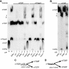
m7GDP, m7GpppG and m7GMP conversion in yeast extracts. (A) Products resulting from the incubation of m7GDP or m7GpppG in various yeast extracts were fractionated by TLC and detected by autoradiography. Purified radiolabeled m7GDP and m7GpppG were incubated with buffer (lanes 1, 4, 7, 10), or whole cell extracts from wild-type yeast (lanes 2, 5, 8, 11) or a Δdcs1 mutant (lanes 3, 6, 9, 12). In lanes 1 to 3 and 7 to 9 addition of ATP was omitted. Lanes labeled M on the left contains molecular markers. Positions of migration of the various cap derivatives are indicated on the left. (B) TLC analysis of m7GMP conversion products. Purified radiolabeled m7GMP was incubated with buffer (lanes 1 and 3), or whole cell extracts from wild-type yeast (lanes 2 and 4). In lanes 1 and 2 addition of ATP was omitted. (C and D) Schemes indicating the fate of m7GDP and m7GpppG in extracts from different strains.
Figure 2.
Figure 2.
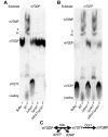
Role of NDK encoded by the YNK1 gene if cap conversion (A). TLC analysis of m7GDP conversion products. Purified radiolabeled m7GDP was incubated with buffer (lanes 1), or whole cell extracts from wild-type yeast (lane 2), a Δdcs1 mutant (lane 3), a Δynk1 mutant (lane 4) or a Δdcs1Δynk1 double mutant (lane 5). (B) Same as in (A) but the substrate is m7GTP. All reactions were performed in presence of ATP. (C) Scheme describing the biochemical pathway involved in m7GDP elimination in extracts.
Figure 3.
Figure 3.
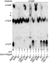
m7GDP conversion in human cell extracts. Products resulting from the incubation of m7GDP in an HEK293 whole-cell extract, or as control yeast extracts, were fractionated by TLC and detected by autoradiography. Purified radiolabeled m7GDP was incubated with buffer (lanes 1, 5 and 9), whole cell extracts from wild-type (lanes 2, 6 and 10) and Δdcs1 (lanes 3, 7 and 11) yeast strains, or HEK293 cell extract (lanes 4, 8, 12). Reactions contained ATP at a final concentration of 2 mM except in lanes 1–4. In lanes 9–12, cold m7GpppG was added to a final concentration of 10 μM to inhibit DcpS. The positions of cap derivatives are indicated on the left side and the left lane labeled M contains molecular markers.
Figure 4.
Figure 4.
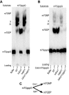
m7GpppG conversion in yeast extracts. A TLC analysis of m7GpppG conversion products is presented. (A). Purified radiolabeled m7GpppG was used as substrate. Products resulting from incubation with buffer (lanes 1), or whole cell extracts from wild-type yeast (lane 2), a Δdcs1 mutant (lane3), a Δaph1 mutant (lane 4) or a Δdcs1Δaph1 mutant (lane 5) were analyzed. (B) Same as in (A) but cold m7GpppG was added to a final concentration of 10 μM to inhibit DcpS.
Figure 5.
Figure 5.
m7GpppG cleavage by recombinant Aph1 and FHIT. (A) Products resulting from the incubation of m7GpppG with recombinant proteins were fractionated by TLC and detected by autoradiography. Purified radiolabeled m7GpppG was incubated with reaction buffer (lane 1), or GST (lane 2), GST-FHIT (lane 3), a GST-FHIT catalytic mutant H96N (lane 4), GST-Aph1 (lane 5), a GST-Aph1 catalytic mutant H109N (lane 6) or as a control human DcpS (lane 7). All reactions contained 0.5 μg of the respective recombinant protein. (B) Purified protein profiles. Recombinant GST:Aph1 and GST:FHIT and the respective catalytic mutants were purified and separated on sodium dodecyl sulphate-polyacrylamide gel electrophoresis. Positions of migration of molecular weight markers are indicated on the right side.
Figure 6.
Figure 6.
m7G cap metabolism in living cells. An artificial 49 nt long capped RNA radiolabeled on the cap phosphate group in position γ was electroporated in cells: wild-type (lane 1), Δynk1 (lane 2), Δdcs1 (lane 3), Δdcs1Δynk1 (lane 4), Δaph1 (lane 5), Δdcs1Δaph1 (lane 6) Δaph1Δynk1 (lane 7) or Δdcs1Δaph1Δynk1 (lane 8) strains. After 45 min of incubation, the resulting metabolites were extracted, fractionated by TLC and detected by autoradiography. Products recovered from the strains above were incubated with 1 unit of NDK in presence of ATP (lanes 9–16) or 2 μg of purified Dcs1 (lanes 17–24). The product resulting from the conversion of species Ypp by NDK (Yppp) is not resolved from m7GTP. The two TLC were developed in parallel and exposed for the same duration.
Figure 7.
Figure 7.
RNA decay pathways and cap (di-)nucleotide elimination mechanisms in yeast and mammalian cells. Enzymes involved in the generation of cap (di-)nucleotide in the 5′-3′ and 3′-5′ mRNA decay pathways are indicated as well as the resulting products. Mechanisms mediating the elimination of these compounds identified in this study are indicated with yeast enzymes indicated in blue and human factors in red. Degradation of the end-product m7GMP by cNIII was reported earlier (46).

Similar articles

Cited by

References

    1. Ghosh A., Lima C.D. Enzymology of RNA cap synthesis. Wiley Interdiscip. Rev. RNA. 2010;1:152–172. - PMC - PubMed
    1. Chan S., Choi E.A., Shi Y. Pre-mRNA 3’-end processing complex assembly and function. Wiley Interdiscip. Rev. RNA. 2011;2:321–335. - PMC - PubMed
    1. Fischer U., Englbrecht C., Chari A. Biogenesis of spliceosomal small nuclear ribonucleoproteins. Wiley Interdiscip. Rev. RNA. 2011;2:718–731. - PubMed
    1. Blumenthal T. Trans-splicing and polycistronic transcription in Caenorhabditis elegans. Trends Genet. 1995;11:132–136. - PubMed
    1. Bangs J.D., Crain P.F., Hashizume T., McCloskey J.A., Boothroyd J.C. Mass spectrometry of mRNA cap 4 from trypanosomatids reveals two novel nucleosides. J. Biol. Chem. 1992;267:9805–9815. - PubMed

Publication types

MeSH terms