

Suitability of antral follicle counts and computer-assisted analysis of ultrasonographic and magnetic resonance images for estimating follicular reserve in porcine, ovine and bovine ovaries ex situ
- PMID: 25432987
- PMCID: PMC4935271
- DOI: 10.1177/1535370214560971
Suitability of antral follicle counts and computer-assisted analysis of ultrasonographic and magnetic resonance images for estimating follicular reserve in porcine, ovine and bovine ovaries ex situ
Abstract
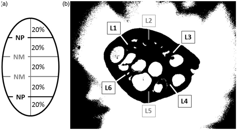
This study was conducted to determine if correlations exist between the numbers of microscopic follicles comprising ovarian follicular reserve (OFR) and antral follicle counts (AFCs), and to assess the usefulness of computerized analyses of ovarian ultrasonograms and magnetic resonance (MR) images for estimating OFR in excised porcine, ovine and bovine ovaries. As a pre-requisite to these analyses, we characterized and compared ovarian cortical histomorphology and follicle populations in the three species varying in prolificacy and overall reproductive longevity, and hence the total number of microscopic and antral follicles. Ultrasonographic and MR images were obtained at the scanner settings optimized to provide opposing contrasts between antral follicles and the ovarian stroma. Commercially available ImageProPlus® analytical software was used to calculate numerical pixel values (NPVs) and pixel heterogeneity (standard deviation of the pixel values) along the computer-generated lines (4-6) placed in the area corresponding to the ovarian cortex. The numbers of primordial (r = 0.38, P < 0.01) and intermediate follicles (r = 0.37, P < 0.01) were correlated with the numbers of antral follicles in bovine ovarian sections. The numbers of primordial (r = 0.28, P < 0.05), intermediate (r = 0.31, P < 0.01) and primary follicles (r = 0.27, P < 0.05) correlated directly with mean NPVs of the ultrasonographic ovarian images in cattle. There was a negative correlation between primary follicle numbers and NPVs of MR images (3D FAST-SPOILED GRADIENT ECHO) of the porcine ovarian cortex (r = -0.31, P < 0.05). To summarize, the numbers of primordial and intermediate follicles could only be estimated from AFCs in cows. Using ultrasound NPVs, the numbers of primordial, intermediate and primary follicles could be directly estimated in bovine ovaries and the quantitative image attributes of MR images were useful for quantifying porcine primary follicles. The bovine ovarian model is compatible with human situation and hence future studies should be undertaken to ascertain the usefulness of AFCs and ultrasonographic image analyses for estimating OFR in women.
Keywords: Ovarian follicle reserve; digital image analysis; magnetic resonance imaging; ovarian histology; ultrasonography.
© 2014 by the Society for Experimental Biology and Medicine.
Figures

Similar articles
-
Relationships between ultrasonographic image attributes, histomorphology and proliferating cell nuclear antigen expression of bovine antral follicles and corpora lutea ex situ.Reprod Domest Anim. 2008 Feb;43(1):27-34. doi: 10.1111/j.1439-0531.2007.00848.x. Reprod Domest Anim. 2008. PMID: 18199255
-
Brangus cows have ovarian reserve parameters more like Brahman than Angus cows.Anim Reprod Sci. 2019 Oct;209:106170. doi: 10.1016/j.anireprosci.2019.106170. Epub 2019 Aug 20. Anim Reprod Sci. 2019. PMID: 31514925
-
Anti-Müllerian hormone inhibits activation and growth of bovine ovarian follicles in vitro and is localized to growing follicles.Mol Hum Reprod. 2017 May 1;23(5):282-291. doi: 10.1093/molehr/gax010. Mol Hum Reprod. 2017. PMID: 28333275 Free PMC article.
-
Antral follicle growth and endocrine changes in prepubertal cattle, sheep and goats.Anim Reprod Sci. 2003 Oct 15;78(3-4):259-70. doi: 10.1016/s0378-4320(03)00094-0. Anim Reprod Sci. 2003. PMID: 12818648 Review.
-
Effects of maternal environment during gestation on ovarian folliculogenesis and consequences for fertility in bovine offspring.Reprod Domest Anim. 2012 Aug;47 Suppl 4:31-7. doi: 10.1111/j.1439-0531.2012.02052.x. Reprod Domest Anim. 2012. PMID: 22827347 Review.
Cited by
-
Platelet-Rich Plasma and Electrochemical Biosensors: A Novel Approach to Ovarian Function Evaluation and Diagnostics.Int J Mol Sci. 2025 Mar 5;26(5):2317. doi: 10.3390/ijms26052317. Int J Mol Sci. 2025. PMID: 40076937 Free PMC article. Review.
-
Serum Concentrations of AMH and E2 and Ovarian and Uterine Traits in Gilts.Animals (Basel). 2019 Oct 15;9(10):811. doi: 10.3390/ani9100811. Animals (Basel). 2019. PMID: 31619004 Free PMC article.
-
A spotlight on factors influencing the in vitro folliculogenesis of isolated preantral follicles.J Assist Reprod Genet. 2024 Dec;41(12):3287-3300. doi: 10.1007/s10815-024-03277-5. Epub 2024 Oct 7. J Assist Reprod Genet. 2024. PMID: 39373807
-
Optimal Dietary Fiber Intake to Retain a Greater Ovarian Follicle Reserve for Gilts.Animals (Basel). 2019 Oct 29;9(11):881. doi: 10.3390/ani9110881. Animals (Basel). 2019. PMID: 31671888 Free PMC article.
-
Ginsenoside Rg1 improves pathological damages by activating the p21‑p53‑STK pathway in ovary and Bax‑Bcl2 in the uterus in premature ovarian insufficiency mouse models.Mol Med Rep. 2021 Jan;23(1):37. doi: 10.3892/mmr.2020.11675. Epub 2020 Nov 12. Mol Med Rep. 2021. PMID: 33179093 Free PMC article.
References
-
- Myers M, Britt KL, Wreford NG, Ebling FJ, Kerr JB. Methods for quantifying follicular numbers within the mouse ovary. Reproduction 2004; 127: 569–80. - PubMed
-
- Kelsey TW, Anderson RA, Wright P, Nelson SM, Wallace WHB. Data-driven assessment of the human ovarian reserve. Mol Hum Reprod 2011; 18: 79–87. - PubMed
-
- Chatterjee S, Modi D, Maitra A, Kadam S, Patel Z, Gokrall J, Meherji P. Screening for FOXL2 gene mutations in women with premature ovarian failure: an Indian experience. Reprod Biomed Online 2007; 15: 554–60. - PubMed
-
- Lass A. Assessment of ovarian reserve: Is there still a role for ovarian biopsy in the light of new data? Hum Reprod 2004; 19: 467–9. - PubMed
Publication types
MeSH terms
LinkOut - more resources
Full Text Sources
Other Literature Sources
Research Materials