. 2015 Feb 1;593(3):521-39.
doi: 10.1113/jphysiol.2014.279109. Epub 2015 Jan 7.

Photoreleasable ligands to study intracrine angiotensin II signalling

Affiliations

Photoreleasable ligands to study intracrine angiotensin II signalling

Artavazd Tadevosyan et al. J Physiol. .

Abstract

Key points: The renin-angiotensin system plays a key role in cardiovascular physiology and its overactivation has been implicated in the pathogenesis of several major cardiovascular diseases. There is growing evidence that angiotensin II (Ang-II) may function as an intracellular peptide to activate intracellular/nuclear receptors and their downstream signalling effectors independently of cell surface receptors. Current methods used to study intracrine Ang-II signalling are limited to indirect approaches because of a lack of selective intracellularly-acting probes. Here, we present novel photoreleasable Ang-II analogues used to probe intracellular actions with spatial and temporal precision. The photorelease of intracellular Ang-II causes nuclear and cytosolic calcium mobilization and initiates the de novo synthesis of RNA in cardiac cells, demonstrating the application of the method.

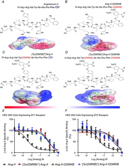
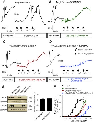
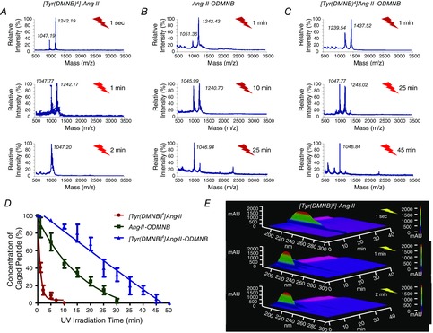
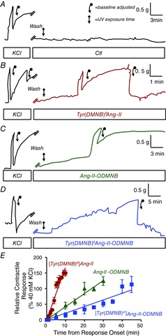
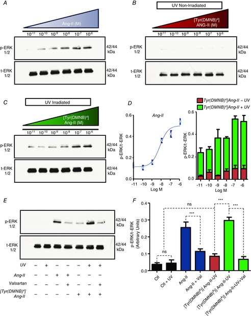
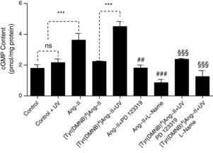
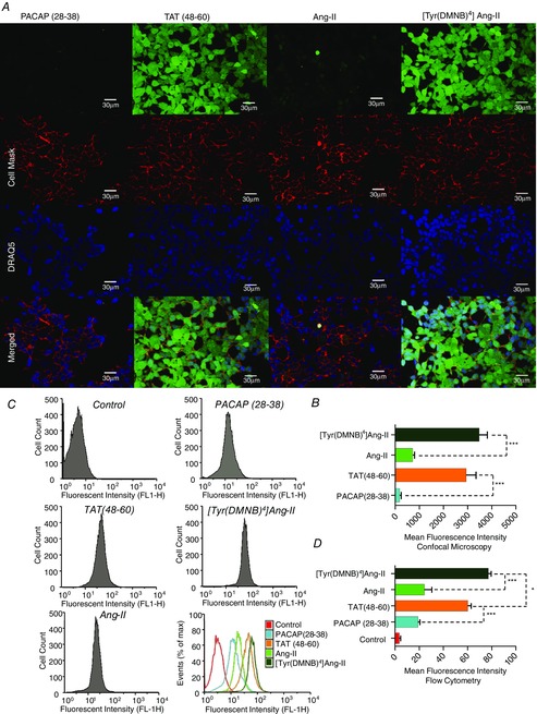
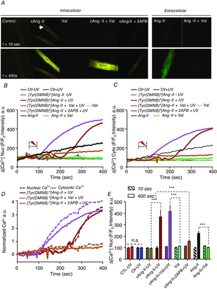
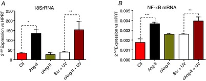
Abstract: Several lines of evidence suggest that intracellular angiotensin II (Ang-II) contributes to the regulation of cardiac contractility, renal salt reabsorption, vascular tone and metabolism; however, work on intracrine Ang-II signalling has been limited to indirect approaches because of a lack of selective intracellularly-acting probes. Here, we aimed to synthesize and characterize cell-permeant Ang-II analogues that are inactive without uncaging, but release active Ang-II upon exposure to a flash of UV-light, and act as novel tools for use in the study of intracrine Ang-II physiology. We prepared three novel caged Ang-II analogues, [Tyr(DMNB)(4)]Ang-II, Ang-II-ODMNB and [Tyr(DMNB)(4)]Ang-II-ODMNB, based upon the incorporation of the photolabile moiety 4,5-dimethoxy-2-nitrobenzyl (DMNB). Compared to Ang-II, the caged Ang-II analogues showed 2-3 orders of magnitude reduced affinity toward both angiotensin type-1 (AT1R) and type-2 (AT2R) receptors in competition binding assays, and greatly-reduced potency in contraction assays of rat thoracic aorta. After receiving UV-irradiation, all three caged Ang-II analogues released Ang-II and potently induced the contraction of rat thoracic aorta. [Tyr(DMNB)(4)]Ang-II showed the most rapid photolysis upon UV-irradiation and was the focus of subsequent characterization. Whereas Ang-II and photolysed [Tyr(DMNB)(4)]Ang-II increased ERK1/2 phosphorylation (via AT1R) and cGMP production (AT2R), caged [Tyr(DMNB)(4)]Ang-II did not. Cellular uptake of [Tyr(DMNB)(4)]Ang-II was 4-fold greater than that of Ang-II and significantly greater than uptake driven by the positive-control HIV TAT(48-60) peptide. Intracellular photolysis of [Tyr(DMNB)(4)]Ang-II induced an increase in nucleoplasmic Ca(2+) ([Ca(2+)]n), and initiated 18S rRNA and nuclear factor kappa B mRNA synthesis in adult cardiac cells. We conclude that caged Ang-II analogues represent powerful new tools for use in the selective study of intracrine signalling via Ang-II.


Figures

Figure 1
Figure 1. Structures of the caged Ang-II analogues and competitive displacement of [125I]Ang-II by caged Ang-II analogues
A, Ang-II. B, Ang-II-ODMNB. C, [Tyr(DMNB)4]Ang-II. D, [Tyr(DMNB)4]Ang-II-ODMNB. The position of the photosensitive DMNB moiety is indicated in brackets and was added either on the side chain of the tyrosine at position 4, the C-terminal carboxylic function of phenylalanine-8, or at both sites. Red and blue surfaces describe negative and positive electrostatic potentials (−3.5 kBT, +3.5 kBT), respectively. Three-dimensional structures were generated using PyMol visualization software. The electrostatic potentials were calculated using the Adaptive Poisson-Boltzmann Solver with the PyMol tool. (E, F, competitive displacement of [125I]Ang-II by Ang-II or caged Ang-II analogues (n = 6/condition) in HEK 293 cells transfected with AT1R or AT2R. Data are percentage of specific radioligand binding in absence of competitors. Non-specific binding was determined in the presence of 1 μm Ang-II.
Figure 2
Figure 2. Concentration-dependent responses of rat thoracic aortic rings to various analogues without uncaging
Contraction-recordings were obtained with a curvilinear pen recorder for (A) Ang-II, (B) Ang-II-ODMNB, (C) [Tyr(DMNB)4]Ang-II and (D) [Tyr(DMNB)4]Ang-II-DMNB. E, immunoblotting for AT1R, AT2R and N-cadherin (positive control) in membranes isolated from rat thoracic aorta, as well as HEK 293, HEK 293-AT1R and HEK 293-AT2R cells. F, overall results expressed as percentage of contractile response induced by 40 mm KCl. Data are shown as the mean ± sem of at least 5 experiments per group performed on tissues isolated from separate animals for each experiment. Descending arrows on original recordings indicate baseline adjustment.
Figure 3
Figure 3. Photolysis kinetics of various analogues
Photolysis (100-W UV-lamp) of a 10−8 m solution of (A) [Tyr(DMNB)4]Ang-II, (B) Ang-II-ODMNB and (C) [Tyr(DMNB)4]Ang-II-ODMNB was performed in Krebs–Henseleit buffer and analysed by mass spectrometry (n = 3 per group). D, caged peptide concentration over time after UV-irradiation. E, signal-intensity (Y-axis) versus wavelength (X-axis) and HPLC elution-time (Z-axis) at different times after photorelease of [Tyr(DMNB)4]Ang-II as analysed by analytical HPLC.
Figure 4
Figure 4. Contractile responses of rat thoracic aortic rings after photolysis of various analogues
Isometric aortic tension recording was obtained with a curvilinear pen recorder after a 15 min incubation in the presence of caged analogues (10−8 m) followed by in situ photolysis with 30-W UV-lamp. A, control. B, [Tyr(DMNB)4]Ang-II. C, Ang-II-ODMNB. D, [Tyr(DMNB)4]Ang-II-DMNB. E, overall results as a percentage of contractile response induced by 40 mm KCl. Data are shown as the mean ± sem (n = 4 independent preparations from each of 4 animals per group).
Figure 5
Figure 5. AT1R-dependent ERK-phosphorylation after photolysis of [Tyr(DMNB)4]Ang-II
Effects of (A) Ang-II, (B) [Tyr(DMNB)4]Ang-II and (C) UV-irradiated [Tyr(DMNB)4]Ang-II on serum starved HEK 293 cells transfected with AT1R. Phosphorylated ERK1/2 (p-ERK) and total ERK1/2 (ERK) immunoreactivity was determined on cell-lysates. D, p-ERK immunoreactivity normalized to total ERK immunoreactivity (mean ± sem). E, p-ERK and ERK immunoreactivity in AT1R-transfected HEK 293 cells treated with vehicle, in the presence or absence of 10 nm Ang-II or [Tyr(DMNB)4]Ang-II (cAng-II), or valsartan (Val; 1 μm, 30 min) with or without UV-irradiation (1 min). F, mean ± sem data corresponding to the experiment in (E) (n = 3 per concentration). **P < 0.01, ***P < 0.001, ns, non-significant.
Figure 6
Figure 6. AT2R-dependent cGMP production after photolysis of [Tyr(DMNB)4]Ang-II
cGMP was measured in serum-starved HEK 293 cells transfected with AT2R after incubation with vehicle (control), Ang-II (10 nm) or [Tyr(DMNB)4]Ang-II, 10 nm) in the presence of PD123319 (PD, 1 μm), l-NAME (1 mm) and UV-irradiation, as indicated. Data are shown as the mean ± sem (n = 3 per condition). ***P < 0.001, ##P < 0.01 or ###P < 0.001 versus Ang-II, §§§P < 0.001 versus [Tyr(DMNB)4]Ang-II. ns, non-significant.
Figure 7
Figure 7. Cellular uptake of fluorescein-[Ahx0,Tyr(DMNB)4]Ang-II
A, intracellular distribution of fluorescein-[Ahx0]Ang-II, fluorescein-[Ahx0,Tyr(DMNB)4]Ang-II, fluorescein-[Ahx0]PACAP(28–38) and fluorescein-[Ahx0]TAT(48–60) upon confocal microscopy in live non-permeabilized HEK 293 cells. Cell Mask and DRAQ5 were used to delineate the plasma membrane and nuclei, respectively. B, intracellular fluorescence intensity (mean ± sem; ***P < 0.001). C, representative uptake efficiency of fluorescein-conjugated PACAP(28–38), TAT(48–60), Ang-II and [Tyr(DMNB)4]Ang-II in HEK 293 cells upon flow cytometry; quantified (D) with fluorescence-activated cell sorting analysis software FlowJo (mean ± sem; n = 3 per condition; *P < 0.05, ***P < 0.001).
Figure 8
Figure 8. Response of nucleoplasmic and cytosolic [Ca2+] to photolysis of intracellular [Tyr(DMNB)4]Ang-II
A, nucleoplasmic and cytosolic [Ca2+] recorded in canine cardiomyocytes before (t = 10 s) and after (t = 400 s) photolysis in cells loaded with vehicle (control), 20 nm [Tyr(DMNB)4]Ang-II (cAng-II), 20 nm cAng-II + 1 μm valsartan, 20 nm cAng-II + 100 μm 2-APB, 1 μm valsartan alone or extracellularly administered 20 nm Ang-II with or without 1 μm valsartan. Photolysis was induced by a pulse of UV-light (2–5 s, 70 μW) from a 405 nm/30 mW diode. Nucleoplasmic (B) and cytosolic (C) [Ca2+] recorded in ventricular cardiomyocytes before and after photolysis in cells preincubated with [Tyr(DMNB)4]Ang-II, vehicle (Ctl), valsartan, 2-APB along with non-photolysed [Tyr(DMNB)4]Ang-II or vehicle. Dotted bracket, UV-irradiation. DRAQ5 fluorescence was used to select the area corresponding to the nucleoplasm. Signals are presented as background-subtracted normalized fluorescence (%F/F0), where F is the fluorescence intensity and F0 is the resting fluorescence in the same cell prior to photolysis. D, [Ca2+] fluorescence in the nucleus and cytosol normalized to respective baseline (t = 10 s) values. Nuclear changes are indicated by dashed lines and cytosolic by continuous lines. E, for each condition (n = 8–12 cells), mean nuclear Fluo-4 fluorescence at baseline (t = 10 s; hatched bars) or after stimulation (t = 400 s) was quantified. Data are shown as the mean ± sem; ***P < 0.000. ns, non-significant.
Figure 9
Figure 9. Photolysis of intracellular [Tyr(DMNB)4]Ang-II regulates transcription
18S rRNA (A) and NF-κB mRNA (B) were quantified by quantitative PCR. A stimulation-control (scr) condition was performed where cardiomyocytes were loaded with a slowly-photolysing Ang-II analogue, Ang-II-ODMNB. Myocytes were incubated with [Tyr(DMNB)4]Ang-II or Ang-II-ODMNB (10 nm) for 30 min at room temperature. After incubation, cells were washed, placed on ice and exposed to a UV-lamp (30 W) for 1 min. In addition, cells were examined after treatment with buffer (Ctl) or extracellular Ang-II (Ang-II; 10 nm). Cells were then incubated at 37°C for 4 h prior to RNA extraction. Data are shown as the mean ± sem; *P < 0.05, **P < 0.01, ***P < 0.0001.

Similar articles

Cited by

References

    1. Ando H, Furuta T, Tsien RY. Okamoto H. Photo-mediated gene activation using caged RNA/DNA in zebrafish embryos. Nat Genet. 2001;28:317–325. - PubMed
    1. Aumelas A, Sakarellos C, Lintner K, Fermandjian S, Khosla MC, Smeby RR. Bumpus FM. Studies on angiotensin II and analogs: impact of substitution in position 8 on conformation and activity. Proc Natl Acad Sci U S A. 1985;82:1881–1885. - PMC - PubMed
    1. Bosnyak S, Jones ES, Christopoulos A, Aguilar MI, Thomas WG. Widdop RE. Relative affinity of angiotensin peptides and novel ligands at AT1 and AT2 receptors. Clin Sci (Lond) 2011;121:297–303. - PubMed
    1. Bourgault S, Letourneau M. Fournier A. Development and pharmacological characterization of "caged" urotensin II analogs. Peptides. 2005;26:1475–1480. - PubMed
    1. Bourgault S, Letourneau M. Fournier A. Development of photolabile caged analogs of endothelin-1. Peptides. 2007;28:1074–1082. - PubMed

Publication types

LinkOut - more resources