. 2015 Feb 1;593(3):585-99.
doi: 10.1113/jphysiol.2014.280792.

Short-chain fatty acid sensing in rat duodenum

Affiliations

Short-chain fatty acid sensing in rat duodenum

Yasutada Akiba et al. J Physiol. .

Abstract

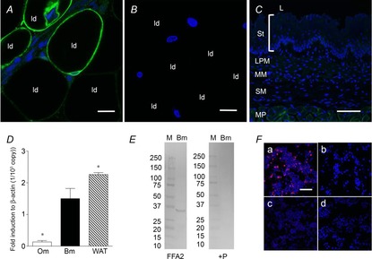
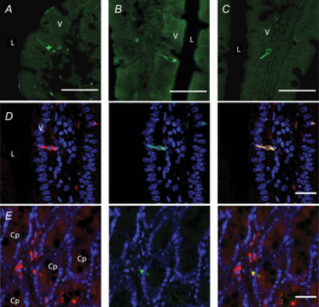
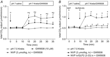
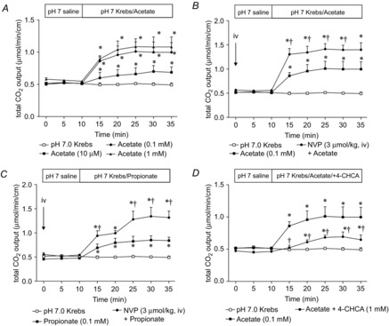
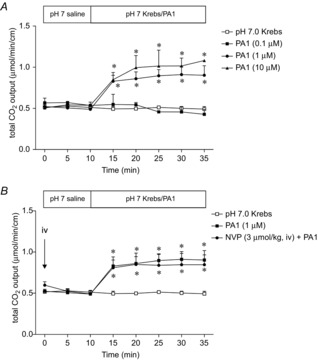
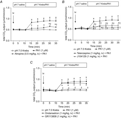
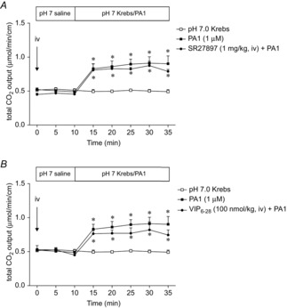
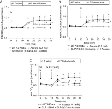
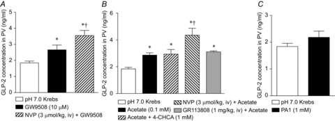
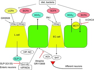
Key points: Luminal lipid in the duodenum modulates gastroduodenal functions via the release of gut hormones and mediators such as cholecystokinin and 5-HT. The effects of luminal short-chain fatty acids (SCFAs) in the foregut are unknown. Free fatty acid receptors (FFARs) for long-chain fatty acids (LCFAs) and SCFAs are expressed in enteroendocrine cells. SCFA receptors, termed FFA2 and FFA3, are expressed in duodenal enterochromaffin cells and L cells, respectively. Activation of LCFA receptor (FFA1) and presumed FFA3 stimulates duodenal HCO3(-) secretion via a glucagon-like peptide (GLP)-2 pathway, whereas FFA2 activation induces HCO3(-) secretion via muscarinic and 5-HT4 receptor activation. The presence of SCFA sensing in the duodenum with GLP-2 and 5-HT signals further supports the hypothesis that luminal SCFA in the foregut may contribute towards the generation of functional symptoms.

Abstract: Intraduodenal fatty acids (FA) and bacterial overgrowth, which generate short-chain FAs (SCFAs), have been implicated in the generation of functional dyspepsia symptoms. We studied the mechanisms by which luminal SCFA perfusion affects duodenal HCO3(-) secretion (DBS), a measure of mucosal neurohumoral activation. Free fatty acid receptor (FFAR) 1 (FFA1), which binds long-chain FA (LCFA), and SCFA receptors FFA2 and FFA3 were immunolocalised to duodenal enteroendocrine cells. FFA3 colocalised with glucagon-like peptide (GLP)-1, whereas FFA2 colocalised with 5-HT. Luminal perfusion of the SCFA acetate or propionate increased DBS, enhanced by dipeptidyl peptidase-IV (DPPIV) inhibition, at the same time as increasing GLP-2 portal blood concentrations. Acetate-induced DBS was partially inhibited by monocarboxylate/HCO3(-) exchanger inhibition without affecting GLP-2 release, implicating acetate absorption in the partial mediation of DBS. A selective FFA2 agonist dose-dependently increased DBS, unaffected by DPPIV inhibition or by cholecystokinin or 5-HT3 receptor antagonists, but was inhibited by atropine and a 5-HT4 antagonist. By contrast, a selective FFA1 agonist increased DBS accompanied by GLP-2 release, enhanced by DPPIV inhibition and inhibited by a GLP-2 receptor antagonist. Activation of FFA1 by LCFA and presumably FFA3 by SCFA increased DBS via GLP-2 release, whereas FFA2 activation stimulated DBS via muscarinic and 5-HT4 receptor activation. SCFA/HCO3(-) exchange also appears to be present in the duodenum. The presence of duodenal fatty acid sensing receptors that signal hormone release and possibly signal neural activation may be implicated in the pathogenesis of functional dyspepsia.


Figures

Figure 1
Figure 1. Detection of FFA2 using RK1101 antibody in rat tissues
Whole mount mesenteric white adipose tissue or a cryostat section of oesophagus was incubated with anti-FFA2 antibody RK1101 (green) with or without blocking peptide, counterstained with 4′,6-diamidino-2-phenylindole (DAPI, blue). Mesenteric adipocytes were positively stained (A), whereas pre-absorption abolished the staining (B). ld, lipid droplet. Internal bars: 20 μm. C, oesophageal mucosa, consisting of stratum layers (St), lamina propria mucosae (LPM), muscularis mucosae (MM) and submucosa (SM), was negatively stained, with faint staining observed in the muscularis propria (MP). L, lumen. Internal bars: 100 μm. D, expression of FFA2 in oesophageal mucosa (Om), duodenal bulb mucosa (Bm) and white adipose tissue (WAT) assessed by real time PCR with the ΔCt method. Each data point represents the mean ± SEM (n = 6 rats). *P < 0.05 vs. Bm. E, Western blot for FFA2 using RK1101 in duodenal mucosa (Bm) (left panel; FFA2) and pre-absorption with blocking peptide (right panel; + P). M, molecular marker with size (kDa) on the left. F, FFA2-transfected cells (a) were positively stained with RK1101 (red), whereas FFA2-transfected cells were negatively stained with pre-absorbed antibody (b). FFA3-transfected cells (c) and mock-transfected cells (d) were negatively stained with RK1101. Counterstained with DAPI (blue). Internal bars: 100 μm.
Figure 2
Figure 2. Localisation of FFARs in rat duodenum
Frozen cryostat sections were incubated with the primary antibody for FFA1 (A), FFA2 (B) or FFA3 (C). L, lumen; V, villus. Internal bars: 100 μm. D, double staining with FFA2 (red; RK1101 antibody, left), 5-HT (green; middle) and merged image (right). Counterstained with DAPI (blue). L, lumen; V, villus. Internal bars: 20 μm. E, double staining with FFA3 (red; left), GLP-1 (green; middle) and merged image (right). Counterstained with DAPI (blue). Cp, crypt. Internal bars: 20 μm.
Figure 3
Figure 3. Effect of FFA1 agonist on duodenal HCO3 secretion in rats
Duodenal HCO3 secretion was measured as total CO2 output using the flow-through pH and CO2 electrodes. The duodenal loop was perfused with GW9508 (10 μm). The DPPIV inhibitor NVP728 (NVP) was injected i.v. (3 μmol kg−1) at t = 0 min (A). The GLP-2 receptor antagonist GLP2(3–33) was additionally injected i.v. (3 nmol kg−1, i.v.) at t = 10 min (B). Each data point represents the mean ± SEM (n = 6 rats). *P < 0.05 vs. pH 7.0 Krebs group. P < 0.05 vs. vehicle i.v. + GW9508 group.
Figure 4
Figure 4. Effect of luminal perfusion of SCFA on duodenal HCO3 secretion
A, luminal perfusion of acetate (10 μm to 1 mm) dose-dependently increased HCO3 secretion. Each data point represents the mean ± SEM (n = 6 rats). *P < 0.05 vs. pH 7.0 Krebs group. B, C, luminal perfusion of acetate (B) or propionate (C) (0.1 mm) increased HCO3 secretion, with the effect enhanced by the DPPIV inhibitor NVP pretreatment (3 μmol kg−1, i.v.). Each data point represents the mean ± SEM (n = 6 rats). *P < 0.05 vs. pH 7.0 Krebs group. P < 0.05 vs. SCFA group. D, co-perfusion of 4-CHCA (1 mm) reduced acetate-induced HCO3 secretion. Each data point represents the mean ± SEM (n = 6 rats). *P < 0.05 vs. pH 7.0 Krebs group. P < 0.05 vs. Acetate group.
Figure 5
Figure 5. Effect of luminal perfusion of FFA2 agonist on HCO3 secretion
A, luminal perfusion of PA1 (0.1–10 μm) dose-dependently increased HCO3 secretion. Each data point represents the mean ± SEM (n = 6 rats). *P < 0.05 vs. pH 7.0 Krebs group. B, PA1 (1 μm)-induced HCO3 secretion was not affected by the DPPIV inhibitor NVP (3 μmol kg−1, i.v.). Each data point represents the mean ± SEM (n = 6 rats). *P < 0.05 vs. pH 7.0 Krebs group.
Figure 6
Figure 6. Effect of muscarinic or 5-HT receptor antagonist on PA1-induced HCO3 secretion
Muscarinic or 5-HT receptor antagonist was injected i.v. at t = 0 min. PA1 (1 μm)-induced HCO3 secretion was inhibited by atropine (0.5 mg kg−1) (A), telenzipine or J104129 (1 mg kg−1) (B). C, 5-HT4 antagonist GR113808 (1 mg kg−1) abolished PA1-induced HCO3 secretion, whereas 5-HT3 receptor antagonist ondansetron (1 mg kg−1) had no effect. Each data point represents the mean ± SEM (n = 6 rats). *P < 0.05 vs. pH 7.0 Krebs group. P < 0.05 vs. PA1 group.
Figure 7
Figure 7. Effect of CCK or VIP receptor antagonist on PA1-induced HCO3 secretion
CCK1 receptor antagonist SR27897 (1 mg kg−1) or VIP receptor antagonist VIP6–28 (100 nmol kg−1) was injected i.v. at t = 0 min. PA1 (1 μm)-induced HCO3 secretion was not affected by SR27897 (A) or VIP6–28 (B). Each data point represents the mean ± SEM (n = 6 rats). *P < 0.05 vs. pH 7.0 Krebs group.
Figure 8
Figure 8. Effect of 5-HT4 receptor or GLP-2 receptor antagonist on acetate-induced HCO3 secretion
GR113808 (1 mg kg−1) was injected i.v. at t = 0 min, whereas GLP-2(3–33) was injected i.v. (3 nmol kg−1, i.v.) at t = 10 min, followed by luminal acetate (0.1 mm) perfusion. GR113808 (A) or GLP-2(3–33) (B) had little effect on acetate-induced HCO3 secretion. Pre-treatment of both antagonists reduced acetate-induced HCO3 secretion (C). Each data point represents the mean ± SEM (n = 4–6 rats). *P < 0.05 vs. pH 7.0 Krebs group. P < 0.05 vs. Acetate group.
Figure 9
Figure 9. Effect of luminal perfusion of fatty acid receptor agonists on GLP-2 release
GLP-2 concentration was measured in PV plasma. The duodenal loop was perfused with GW9508 (10 μm, A), acetate (0.1 mm, B) or PA1 (1 μm, C) with or without inhibitors. PV blood was collected at t = 35 min. Each data point represents the mean ± SEM (n = 6 rats). *P < 0.05 vs. pH 7.0 Krebs group.
Figure 10
Figure 10. Proposed mechanism of duodenal SCFA sensing
Luminal SCFAs, derived from diet or bacterial metabolism, activate multiple pathways. SCFAs may activate FFA3 on L cells, which release GLP-2, which in turn activates GLP-2 receptors expressed on myenteric neurons (Guan et al. 2006), followed by the release of VIP and NO (Wang et al. 2011), stimulating epithelial HCO3 secretion by established pathways. The actions of GLP-2 are enhanced by DPPIV inhibition (Inoue et al. 2012). SCFAs also activate FFA2 on EC cells, which release 5-HT and ACh, activating 5-HT4 and muscarinic receptors, respectively, with both expressed on enteric and afferent nerves, and on epithelial cells, also stimulating the rate of HCO3 secretion. SCFAs might be absorbed by SCFA/HCO3 exchanger, possibly by apical membrane MCT1, increasing the rate of HCO3 secretion. By contrast, luminal LCFA increases HCO3 secretion via activation of FFA1 expressed in L cells, which releases GLP-2, augmented by DPPIV inhibition, and inhibited by a GLP-2 receptor antagonist GLP-2(3–33). We propose that GLP-2, 5-HT and ACh, which are released by luminal SCFAs, not only activate the local mechanisms involved in duodenal mucosal defences, but also stimulate afferent nerves, which are implicated in the production of pathological symptoms. SCFA, short-chain fatty acid; LCFA, long-chain fatty acid; FFA1, free fatty acid receptor 1; GLP-2, glucagon-like peptide-2; VIP, vasoactive intestinal peptide; NO, nitric oxide; 5-HT, 5-hydroxytryptamine; Ach, acetylcholine; EC, enterochromaffin; MCT, monocarboxylate transporter; DPPIV, dipeptidyl peptidase IV.

References

    1. Akiba Y. Kaunitz JD. Duodenal chemosensing and mucosal defenses. Digestion. 2011;83(Suppl 1):25–31. - PMC - PubMed
    1. Akiba Y. Kaunitz JD. Duodenal chemosensing: Master control for epigastric sensation. J Gastroenterol Hepatol. 2011;26:6–7. - PubMed
    1. Akiba Y, Mizumori M, Guth PH, Engel E. Kaunitz JD. Duodenal brush border intestinal alkaline phosphatase activity affects bicarbonate secretion in rats. Am J Physiol Gastrointest Liver Physiol. 2007;293:G1223–G1233. - PubMed
    1. Akiba Y, Mizumori M, Kuo M, Ham M, Guth PH, Engel E. Kaunitz JD. CO2 chemosensing in rat oesophagus. Gut. 2008;57:1654–1664. - PMC - PubMed
    1. Akiba Y, Watanabe C, Mizumori M. Kaunitz JD. Luminal L-glutamate enhances duodenal mucosal defense mechanisms via multiple glutamate receptors in rats. Am J Physiol Gastrointest Liver Physiol. 2009;297:G781–G791. - PMC - PubMed

Publication types

LinkOut - more resources