

Microglia inflammatory responses are controlled by an intrinsic circadian clock
- PMID: 25433170
- PMCID: PMC4386638
- DOI: 10.1016/j.bbi.2014.11.009
Microglia inflammatory responses are controlled by an intrinsic circadian clock
Abstract
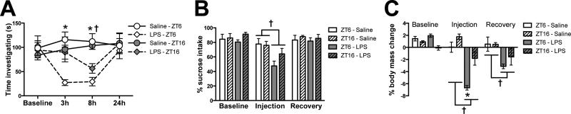
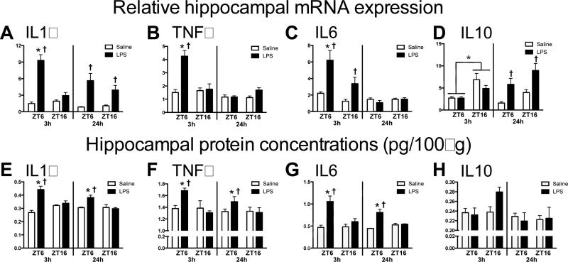
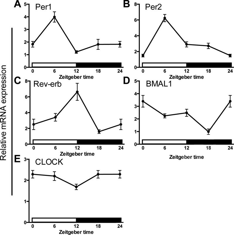
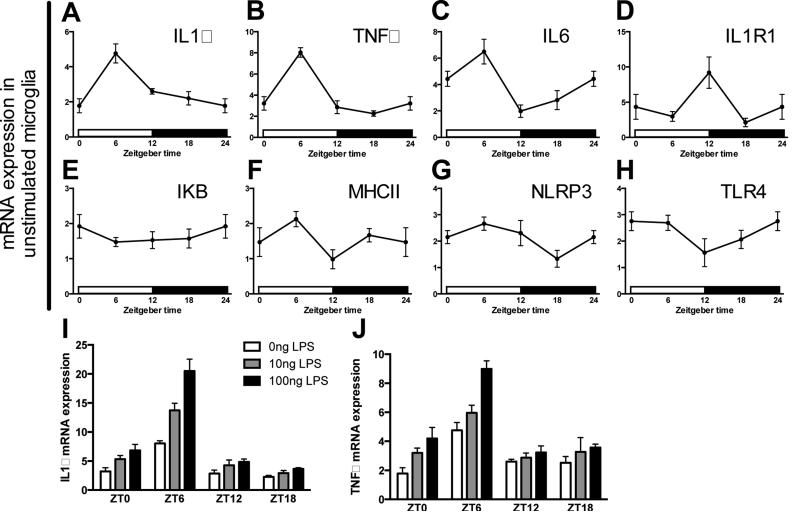
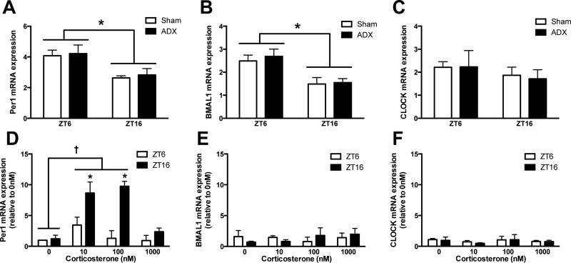
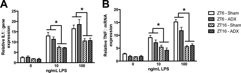
The circadian system regulates many physiological functions including inflammatory responses. For example, mortality caused by lipopolysaccharide (LPS) injection varies depending on the time of immunostimulation in mammals. The effects of more subtle challenges on the immune system and cellular mechanisms underlying circadian differences in neuroinflammatory responses are not well understood. Here we show that adult male Sprague-Dawley rats injected with a sub-septic dose of LPS during the light phase displayed elevated sickness behaviors and hippocampal cytokine production compared to rats injected during the dark phase. Microglia are the primary central nervous system (CNS) immune cell type and may mediate diurnal differences in sickness response, thus we explored whether microglia demonstrate temporal variations in inflammatory factors. Hippocampal microglia isolated from adult rats rhythmically expressed inflammatory factors and circadian clock genes. Microglia displayed robust rhythms of TNFα, IL1β and IL6 mRNA, with peak cytokine gene expression occurring during the middle of the light phase. Microglia isolated during the light phase were also more reactive to immune stimulation; such that, ex vivo LPS treatment induced an exaggerated cytokine response in light phase-isolated microglia. Treating microglia with corticosterone ex vivo induced expression of the circadian clock gene Per1. However, microglia isolated from adrenalectomized rats maintained temporal differences in clock and inflammatory gene expression. This suggests circadian clock gene expression in microglia is entrained by, but oscillates in the absence of, glucocorticoids. Taken together, these findings demonstrate that microglia possess a circadian clock that influences inflammatory responses. These results indicate time-of-day is an important factor to consider when planning inflammatory interventions such as surgeries or immunotherapies.
Keywords: Circadian; Clock gene; Cytokines; Glucocorticoids; Inflammation; Microglia.
Copyright © 2014 Elsevier Inc. All rights reserved.
Figures
Similar articles
-
Stress-induced neuroinflammatory priming is time of day dependent.Psychoneuroendocrinology. 2016 Apr;66:82-90. doi: 10.1016/j.psyneuen.2016.01.006. Epub 2016 Jan 11. Psychoneuroendocrinology. 2016. PMID: 26799851 Free PMC article.
-
Diminished circadian rhythms in hippocampal microglia may contribute to age-related neuroinflammatory sensitization.Neurobiol Aging. 2016 Nov;47:102-112. doi: 10.1016/j.neurobiolaging.2016.07.019. Epub 2016 Aug 1. Neurobiol Aging. 2016. PMID: 27568094 Free PMC article.
-
Glucocorticoids mediate stress-induced priming of microglial pro-inflammatory responses.Brain Behav Immun. 2012 Feb;26(2):337-45. doi: 10.1016/j.bbi.2011.10.005. Epub 2011 Oct 24. Brain Behav Immun. 2012. PMID: 22041296 Free PMC article.
-
Chronopharmacological strategies focused on chrono-drug discovery.Pharmacol Ther. 2019 Oct;202:72-90. doi: 10.1016/j.pharmthera.2019.05.018. Epub 2019 Jun 5. Pharmacol Ther. 2019. PMID: 31173839 Review.
-
Differences in Diurnal Variation of Immune Responses in Microglia and Macrophages: Review and Perspectives.Cell Mol Neurobiol. 2020 Apr;40(3):301-309. doi: 10.1007/s10571-019-00736-x. Epub 2019 Sep 23. Cell Mol Neurobiol. 2020. PMID: 31549296 Free PMC article. Review.
Cited by
-
Sleep and Memory Consolidation Dysfunction in Psychiatric Disorders: Evidence for the Involvement of Extracellular Matrix Molecules.Front Neurosci. 2021 May 14;15:646678. doi: 10.3389/fnins.2021.646678. eCollection 2021. Front Neurosci. 2021. PMID: 34054408 Free PMC article. Review.
-
Inflammaging and Brain Aging.Int J Mol Sci. 2024 Sep 30;25(19):10535. doi: 10.3390/ijms251910535. Int J Mol Sci. 2024. PMID: 39408862 Free PMC article. Review.
-
Integration of circadian rhythms and immunotherapy for enhanced precision in brain cancer treatment.EBioMedicine. 2024 Nov;109:105395. doi: 10.1016/j.ebiom.2024.105395. Epub 2024 Oct 15. EBioMedicine. 2024. PMID: 39413708 Free PMC article. Review.
-
The Role of Circadian Rhythms in Stroke: A Narrative Review.Neurochem Res. 2024 Feb;49(2):290-305. doi: 10.1007/s11064-023-04040-5. Epub 2023 Oct 14. Neurochem Res. 2024. PMID: 37838637 Review.
-
The Circadian Molecular Machinery in CNS Cells: A Fine Tuner of Neuronal and Glial Activity With Space/Time Resolution.Front Mol Neurosci. 2022 Jul 1;15:937174. doi: 10.3389/fnmol.2022.937174. eCollection 2022. Front Mol Neurosci. 2022. PMID: 35845604 Free PMC article. Review.
References
-
- Arjona A, Sarkar DK. Evidence supporting a circadian control of natural killer cell function. Brain Behav Immun. 2006;20:469–476. - PubMed
-
- Balsalobre A, Brown SA, Marcacci L, Tronche F, Kellendonk C, Reichardt HM, Schutz G, Schibler U. Resetting of circadian time in peripheral tissues by glucocorticoid signaling. Science. 2000;289:2344–2347. - PubMed
-
- Boivin DB, James FO, Wu A, Cho-Park PF, Xiong H, Sun ZS. Circadian clock genes oscillate in human peripheral blood mononuclear cells. Blood. 2003;102:4143–4145. - PubMed
Publication types
MeSH terms
Substances
Grants and funding
LinkOut - more resources
Full Text Sources
Other Literature Sources
Medical
