

Improvement of the skeletal and dental hypophosphatasia phenotype in Alpl-/- mice by administration of soluble (non-targeted) chimeric alkaline phosphatase
- PMID: 25433339
- PMCID: PMC4283789
- DOI: 10.1016/j.bone.2014.11.017
Improvement of the skeletal and dental hypophosphatasia phenotype in Alpl-/- mice by administration of soluble (non-targeted) chimeric alkaline phosphatase
Abstract
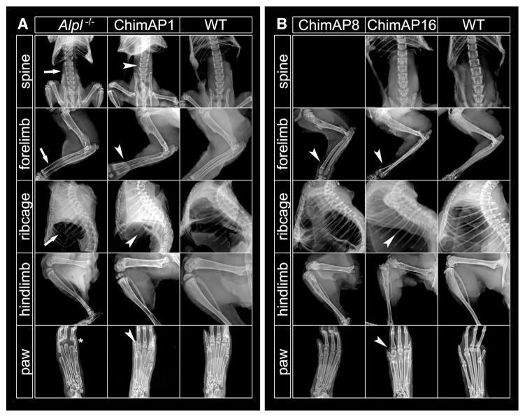
Hypophosphatasia (HPP) results from ALPL gene mutations, which lead to a deficiency of tissue-nonspecific alkaline phosphatase (TNAP), and accumulation of inorganic pyrophosphate, a potent inhibitor of mineralization that is also a natural substrate of TNAP, in the extracellular space. HPP causes mineralization disorders including soft bones (rickets or osteomalacia) and defects in teeth and periodontal tissues. Enzyme replacement therapy using mineral-targeting recombinant TNAP has proven effective in preventing skeletal and dental defects in TNAP knockout (Alpl(-/-)) mice, a model for life-threatening HPP. Here, we show that the administration of a soluble, intestinal-like chimeric alkaline phosphatase (ChimAP) improves the manifestations of HPP in Alpl(-/-) mice. Mice received daily subcutaneous injections of ChimAP at doses of 1, 8 or 16 mg/kg, from birth for up to 53 days. Lifespan and body weight of Alpl(-/-) mice were normalized, and vitamin B6-associated seizures were absent with 16 mg/kg/day of ChimAP. Radiographs, μCT and histological analyses documented improved mineralization in cortical and trabecular bone and secondary ossification centers in long bones of ChimAP16-treated mice. There was no evidence of craniosynostosis in the ChimAP16-treated mice and we did not detect ectopic calcification by radiography and histology in the aortas, stomachs, kidneys or lungs in any of the treatment groups. Molar tooth development and function improved with the highest ChimAP dose, including enamel, dentin, and tooth morphology. Cementum remained deficient and alveolar bone mineralization was reduced compared to controls, though ChimAP-treated Alpl(-/-) mice featured periodontal attachment and retained teeth. This study provides the first evidence for the pharmacological efficacy of ChimAP for use in the treatment of skeletal and dental manifestations of HPP.
Keywords: Craniosynostosis; Enzyme replacement; Hypophosphatasia; Osteomalacia; Rickets; Seizures.
Copyright © 2014 Elsevier Inc. All rights reserved.
Figures


Similar articles
-
Conditional Alpl Ablation Phenocopies Dental Defects of Hypophosphatasia.J Dent Res. 2017 Jan;96(1):81-91. doi: 10.1177/0022034516663633. Epub 2016 Oct 1. J Dent Res. 2017. PMID: 27582029 Free PMC article.
-
Gene Therapy Using Adeno-Associated Virus Serotype 8 Encoding TNAP-D10 Improves the Skeletal and Dentoalveolar Phenotypes in Alpl-/- Mice.J Bone Miner Res. 2021 Sep;36(9):1835-1849. doi: 10.1002/jbmr.4382. Epub 2021 Jun 15. J Bone Miner Res. 2021. PMID: 34076297 Free PMC article.
-
Periodontal Defects in the A116T Knock-in Murine Model of Odontohypophosphatasia.J Dent Res. 2015 May;94(5):706-14. doi: 10.1177/0022034515573273. Epub 2015 Feb 25. J Dent Res. 2015. PMID: 25716980 Free PMC article.
-
Alkaline Phosphatase and Hypophosphatasia.Calcif Tissue Int. 2016 Apr;98(4):398-416. doi: 10.1007/s00223-015-0079-1. Epub 2015 Nov 21. Calcif Tissue Int. 2016. PMID: 26590809 Free PMC article. Review.
-
Dental manifestations of hypophosphatasia: translational and clinical advances.JBMR Plus. 2025 Jan 6;9(2):ziae180. doi: 10.1093/jbmrpl/ziae180. eCollection 2025 Feb. JBMR Plus. 2025. PMID: 39872235 Free PMC article. Review.
Cited by
-
Conditional Alpl Ablation Phenocopies Dental Defects of Hypophosphatasia.J Dent Res. 2017 Jan;96(1):81-91. doi: 10.1177/0022034516663633. Epub 2016 Oct 1. J Dent Res. 2017. PMID: 27582029 Free PMC article.
-
Prevention of Lethal Murine Hypophosphatasia by Neonatal Ex Vivo Gene Therapy Using Lentivirally Transduced Bone Marrow Cells.Hum Gene Ther. 2015 Dec;26(12):801-12. doi: 10.1089/hum.2015.078. Epub 2015 Nov 19. Hum Gene Ther. 2015. PMID: 26467745 Free PMC article.
-
Transcriptional Regulation of Dental Epithelial Cell Fate.Int J Mol Sci. 2020 Nov 25;21(23):8952. doi: 10.3390/ijms21238952. Int J Mol Sci. 2020. PMID: 33255698 Free PMC article. Review.
-
Alkaline Phosphatase Replacement Therapy for Hypophosphatasia in Development and Practice.Adv Exp Med Biol. 2019;1148:279-322. doi: 10.1007/978-981-13-7709-9_13. Adv Exp Med Biol. 2019. PMID: 31482504 Review.
-
Identification of Biomarkers for Osteosarcoma Based on Integration Strategy.Med Sci Monit. 2020 Mar 16;26:e920803. doi: 10.12659/MSM.920803. Med Sci Monit. 2020. PMID: 32173717 Free PMC article.
References
-
- Anderson HC, Harmey D, Camacho NP, Garimella R, Sipe JB, Tague S, et al. Sustained osteomalacia of long bones despite major improvement in other hypophosphatasia-related mineral deficits in tissue nonspecific alkaline phosphatase/nucleotide pyrophosphatase phosphodiesterase 1 double-deficient mice. Am J Pathol. 2005;166(6):1711–20. - PMC - PubMed
-
- Baumgartner-Sigl S, Haberlandt E, Mumm S, Scholl-Bürgi S, Sergi C, Ryan L, et al. Pyridoxine-responsive seizures as the first symptom of infantile hypophosphatasia caused by two novel missense mutations (c.677T>C, p.M226T; c.1112C>T, p. T371I) of the tissue-nonspecific alkaline phosphatase gene. Bone. 2007;40:1655–61. - PubMed
-
- Bouxsein ML, Boyd SK, Christiansen BA, Guldberg RE, Jepsen KJ, Müller R. Guidelines for assessment of bone microstructure in rodents using micro-computed tomography. J Bone Miner Res. 2010;25(7):1468–86. - PubMed
-
- Cahill RA, Wenkert D, Perlman SA, Steele A, Coburn SP, Mcalister WH, et al. Infantile hypophosphatasia: transplantation therapy trial using bone fragments and cultured osteoblasts. J Clin Endocrinol Metab. 2007;92:2923–30. - PubMed
Publication types
MeSH terms
Substances
Grants and funding
LinkOut - more resources
Full Text Sources
Other Literature Sources
Molecular Biology Databases
Miscellaneous
