. 2015 Jan 2;48(1):113-21.
doi: 10.1016/j.jbiomech.2014.10.031. Epub 2014 Nov 1.

Myh11(R247C/R247C) mutations increase thoracic aorta vulnerability to intramural damage despite a general biomechanical adaptivity

Affiliations

Myh11(R247C/R247C) mutations increase thoracic aorta vulnerability to intramural damage despite a general biomechanical adaptivity

Chiara Bellini et al. J Biomech. .

Abstract

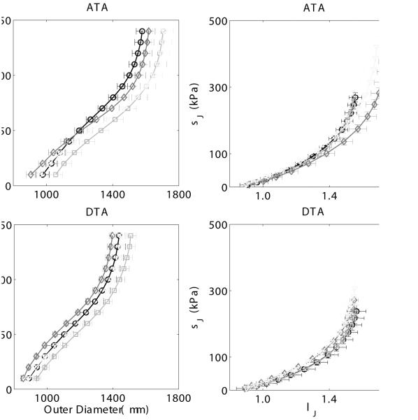
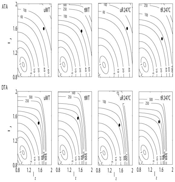
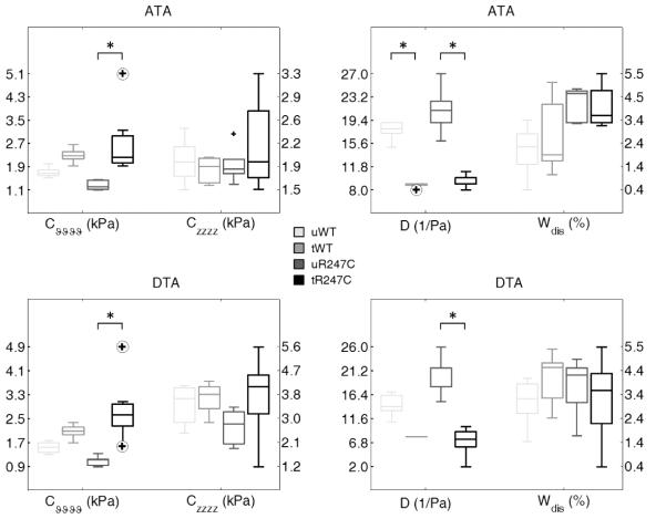
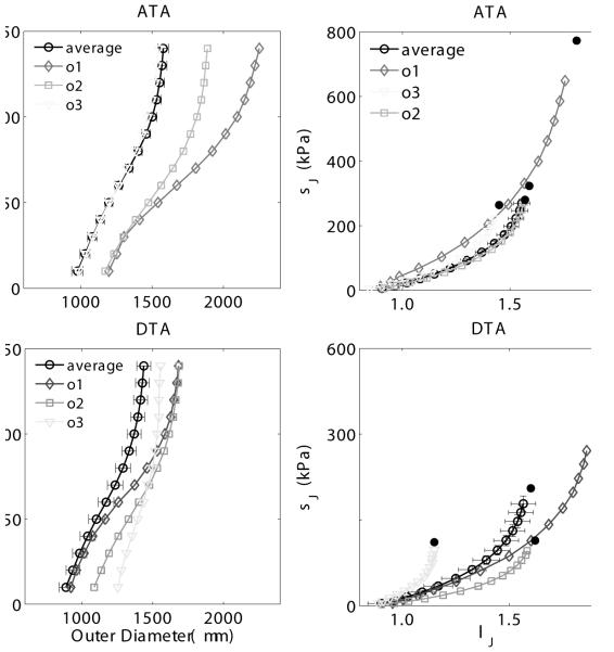
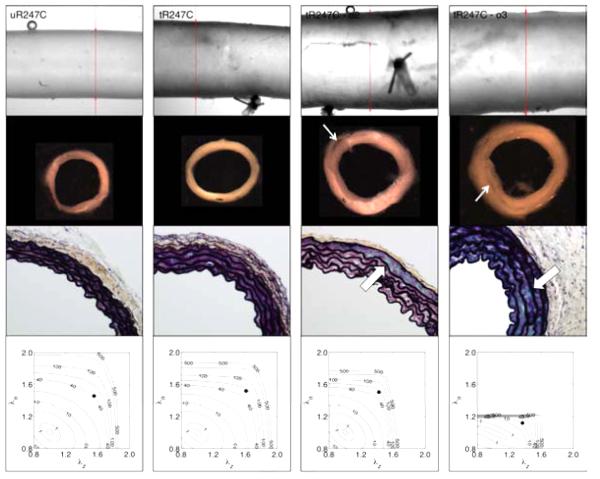
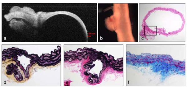
Genetic studies in patients reveal that mutations to genes that encode contractile proteins in medial smooth muscle cells can cause thoracic aortic aneurysms and dissections. Mouse models of such mutations, including Acta2(-/-) and Myh11(R247C/R247C), surprisingly do not present with any severe vascular phenotype under normal conditions. This observation raises the question whether these mutations nevertheless render the thoracic aorta increasingly vulnerable to aneurysms or dissections in the presence of additional, epigenetic, factors such as hypertension, a known risk factor for thoracic aortic disease. Accordingly, we compared the structure and biaxial mechanical properties of the ascending and descending thoracic aorta from male wild-type and Myh11(R247C/R247C) mice under normotension and induced hypertension. On average, the mutant aortas exhibited near normal biomechanics under normotensive hemodynamics and near normal adaptations to hypertensive hemodynamics, yet the latter led to intramural delaminations or premature deaths in over 20% of these mice. Moreover, the delaminated vessels exhibited localized pools of mucoid material, similar to the common histopathologic characteristic observed in aortas from humans affected by thoracic aortic aneurysms and dissections. The present findings suggest, therefore, that mutations to smooth muscle cell contractile proteins may place the thoracic aorta at increased risk to epigenetic factors and that there is a need to focus on focal, not global, changes in aortic structure and properties, including the pooling of glycosaminoglycans/proteoglycans that may lead to thoracic aortic dissection.

Keywords: Actomyosin functionality; Familial thoracic aortic aneurysms and dissections; Smooth muscle myosin heavy chain; Wall stress and stiffness.


Figures

Figure 1
Figure 1
Mean ± SEM pressure - diameter responses (left panels) and circumferential Cauchy stress - stretch responses (right panels) for the ascending thoracic aorta (ATA, top panels) and the proximal descending thoracic aorta (DTA, bottom panels), excluding data from the 4 outliers. Datasets for the untreated (u) and treated (t, L-NAME + NaCl) wild-type (WT) as wells as for untreated (u) and treated (t) Myh11 R247C/R247C (R247C) mice reveal only modest differences due to either the mutation or the induced hypertension (i.e., L-NAME plus high salt treatment). Mean P-d curves were obtained by averaging 5 ATAs/DTAs for the uWT, the uR247C, and the tR247C groups, and 4 ATAs/DTAs for the tWT group.
Figure 2
Figure 2
Contour plots of constant strain energy (W) as a function of biaxial stretch λθ - λz for the untreated wild-type (first column), treated wild-type (second column), untreated Myh11 R247C/R247C (third column) and treated Myh11 R247C/R247C (fourth column) mice. The strain energy functions were estimated from average datasets for the ascending thoracic aorta (ATA, top panels) and proximal descending thoracic aorta (DTA, bottom panels). The solid black circle in each plot indicates the in vivo conditions (mean arterial pressure and preferred axial stretch). The superimposed numerical values on the contours denote the values of strain energy in kPa. The mean strain energy functions (W) representative for each combination of genotype, treatment, and location were obtained by averaging experimental data from 5 ATAs/DTAs for the uWT, the uR247C, and the tR247C groups, and 4 ATAs/DTAs for the tWT group.
Figure 3
Figure 3
Mechanical metrics of material stiffness (left panels), and structural stiffness and energy dissipation (rigth panels) for the ascending thoracic aorta (ATA, top panels) and proximal descending thoracic aorta (DTA, bottom panels) of untreated (u) and treated (L-Name + NaCl, t) wild-type (WT) as well as for untreated (u) and treated (t) Myh11R247C/R247C (R247C) mice, showed at in vivo conditions (mean arterial pressure and preferred axial stretch). Cvvvv and Czzzz are values of linearized stiffness in circumferential and axial direction, respectively. D is the distensibility computed at in vivo conditions (mean arterial pressure and preferred axial stretch). Wdis is the percentage of energy dissipated during the mechanical test, relative to the energy stored after loading. Note: circled cross symbols indicate outliers. Data from all collected vessels were included in the statistical analysis, i.e. 5 ATAs/DTAs for the uWT, tWT, and the uR247C groups, 7 ATAs and 8 DTAs for the tR247C group.
Figure 4
Figure 4
Pressure - diameter (left panels) and circumferential stress - stretch (rigth panels) data for the ascending thoracic aorta (ATA, top panels) and the proximal descending thoracic aorta (DTA, bottom panels). The datasets corresponding to the 3 treated Myh11 R247C/R247C mutant outliers (ol, o2, o3) are compared with the average dataset for the treated mutant group in each plot, for both ATA and DTAs. The standard error of the mean is shown for selected points of the average dataset. Mean P-d curves for the tR247C group were obtained by averaging the data from the 5 ATAs and the 5 DTAs (5 treated mutant mice) that were not classified as outliers.
Figure 5
Figure 5
Gross appearance, microstructure and mechanical behavior of descending thoracic aortas from one representative ‘average’ untreated Myh11 R247C/R247C mouse (first column, uR247C), one representative ‘average’ treated Myh11 R247C/R247C mouse (second column, tR247C), and two of the three treated Myh11 R247C/R247C 'outlier’ mice (third and fourth columns, tR247C-o2 and tR247C-o3 ). The panels in the first row show the gross appearance during biaxial testing, the panels in the second row show cross-sections of rings cut from the vessel after mechanical testing, the panels in the third row show Movat-stained histological sections (60x magnification), and the panels in the forth row show the contour plots of constant strain energy (W). The thin white arrows highlight intramural delaminations that were evident by eye. The thick white arrows highlight accumulated mucoid material, which tended to increase diffusely with induced hypertension but to accumulate more focally in the outliers having evidence of intramural delaminations. Note that the histological sections were necessarily taken from different regions than the isolated rings. Similar to Figure 3, the solid black circle in each contour plot indicates the specimen-specific in vivo condition (mean arterial pressure and preferred axial stretch). The superimposed numerical values on the contours denote the values of strain energy in kPa.
Figure 6
Figure 6
Full vessel and stained cross-sectional views of the proximal descending thoracic aorta from the second outlier in the Myh11 R247C/R247C treated group. A dense mesh of fibrotic tissue originated from the adventitial layer of the aorta (b, naked eye), in the proximity of one of the intercostal branches (a, Optical Coherence Tomographic image during mechanical testing). Cross-sectional cuts stained with H&E (c, lOx magnification), Movat (d, 40x magnification), VVG (e, 40× magnification), and MTC (f, 40× magnification) selectively show cells and/or ECM components and reveal a separation between the elastic laminae within the fibrotic region (magnified).

References

    1. Bersi MR, Ferruzzi J, Eberth JF, Gleason RL, Jr, Humphrey JD. Consistent biomechanical phenotyping of common carotid arteries from seven genetic, pharmacological, and surgical mouse models. Ann Biomed Eng. 2014 Jun;42(6):1207–23. - PMC - PubMed
    1. Chan KK, Rabkin SW. Increasing prevalence of hypertension among patients with thoracic aorta dissection: Trends over eight decades-a structured meta-analysis. Am J Hypertens. 2014 Jul;27(7):907–17. - PubMed
    1. Chen J, Wu J, Li L, Zou Y-Z, Zhu D-L, Gao P-J. Effect of an acute mechanical stimulus on aortic structure in the transverse aortic constriction mouse model. Clin Exp Pharmacol Physiol. 2011 Sep;38(9):570–6. - PubMed
    1. de Wit A, Vis K, Jeremy RW. Aortic stiffness in heritable aortopathies: relationship to aneurysm growth rate. Heart Lung Circ. 2013 Jan;22(1):3–11. - PubMed
    1. El-Hamamsy I, Yacoub MH. Cellular and molecular mechanisms of thoracic aortic aneurysms. Nat Rev Cardiol. 2009 Dec;6(12):771–86. - PubMed

Publication types