. 2014 Nov 30:14:280.
doi: 10.1186/s12866-014-0280-9.

Investigating the genetic regulation of the ECF sigma factor σS in Staphylococcus aureus

Investigating the genetic regulation of the ECF sigma factor σS in Staphylococcus aureus

Whittney N Burda et al. BMC Microbiol. .

Abstract

Background: We previously identified an ECF sigma factor, σS, that is important in the stress and virulence response of Staphylococcus aureus. Transcriptional profiling of sigS revealed that it is differentially expressed in many laboratory and clinical isolates, suggesting the existence of regulatory networks that modulates its expression.

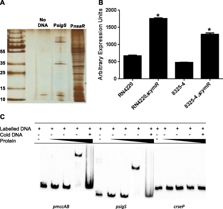
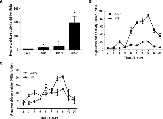
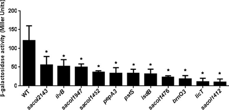
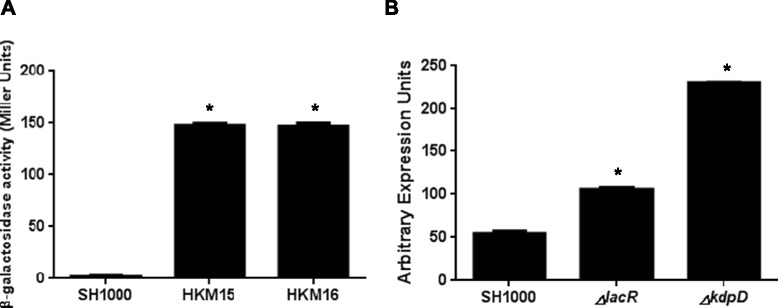
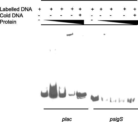
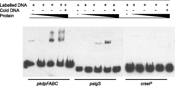
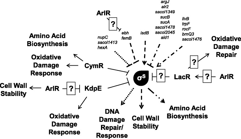
Results: To identify regulators of sigS, we performed a pull down assay using S. aureus lysates and the sigS promoter. Through this we identified CymR as a negative effector of sigS expression. Electrophoretic mobility shift assays (EMSAs) revealed that CymR directly binds to the sigS promoter and negatively effects transcription. To more globally explore genetic regulation of sigS, a Tn551 transposon screen was performed, and identified insertions in genes that are involved in amino acid biosynthesis, DNA replication, recombination and repair pathways, and transcriptional regulators. In efforts to identify gain of function mutations, methyl nitro-nitrosoguanidine mutagenesis was performed on a sigS-lacZ reporter fusion strain. From this a number of clones displaying sigS upregulation were subject to whole genome sequencing, leading to the identification of the lactose phosphotransferase repressor, lacR, and the membrane histidine kinase, kdpD, as central regulators of sigS expression. Again using EMSAs we determined that LacR is an indirect regulator of sigS expression, while the response regulator, KdpE, directly binds to the promoter region of sigS.

Conclusions: Collectively, our work suggests a complex regulatory network exists in S. aureus that modulates expression of the ECF sigma factor, σS.


Figures

Figure 1
Figure 1
CymR is a direct repressor of transcription from the sigS promoter. (A) A pull down assay was performed using crude protein lysates harvested from RN4220 at hour 2 and a biotinylated sigS promoter DNA probe. The identity of protein bands was determined by LC/MS analysis. (B) qPCR analysis of sigS expression in a cymR mutant compared to its respective parental strain, in the 8325–4 and RN4220 backgrounds. Error bars are shown as ± SEM, * = p <0.05 using a Student’s t-test. (C) An electrophoretic mobility shift assay was performed using purified CymR, and the promoter region of mccAB (positive control), the promoter region of sigS (test), and an intergenic region from the rseP gene (rseP; negative control). CymR was added at increasing concentrations of 0.01, 0.1 and 1 μM in all panels.
Figure 2
Figure 2
Transcriptional profiling of sigS in transposon mutants found to negatively effect expression. (A, B, and C) Mutant strains bearing a sigS-lacZ fusion were grown in TSB at 37°C and samples withdrawn at the times specified. β-Galactosidase activity was measured using 4-MUG as a substrate to determine sigS expression levels. Assays were performed on duplicate samples and the values averaged. The results presented are from three independent experiments. Error bars are shown as ± SEM. Significance was determined by a Student t test; *indicates a p value of <0.05.
Figure 3
Figure 3
Identification of positive regulators of sigS expression. Mutant strains bearing a sigS-lacZ fusion were grown in TSB at 37°C and sampled after 5 h of growth. β-Galactosidase activity was measured using 4-MUG as a substrate to determine sigS expression levels. Assays were performed on duplicate samples and the values averaged. The results presented are from three independent experiments. Error bars are shown as ± SEM. Significance was determined using a Student t test; *indicates a p value of <0.05.
Figure 4
Figure 4
Nitrosoguanidine mutagenesis identifies additional regulators of sigS expression. (A) The SH1000 sigS-lacZ fusion strain was subjected to MNNG mutagenesis. Two resulting clones were selected for further analysis during growth in TSB at 37°C. HKM15 and HKM16 were grown along side the parent strain, SH1000, with samples collected at 5 hours. β-Galactosidase activity was measured using 4-MUG as a substrate to determine sigS expression levels. Assays were performed on duplicate samples and the values averaged. The results presented are from three independent experiments. Error bars are shown as ± SEM. (B) qPCR for sigS expression was performed on SH1000 strains containing a mutation in either lacR or kdpD. Error bars are shown as ± SEM. Significance was determined using a Student t test; *indicates a p value of <0.05.
Figure 5
Figure 5
LacR does not directly regulate sigS expression. Electrophoretic mobility shift mobility assays were performed using purified LacR, the lac promoter region (positive control), and the promoter region of sigS (test). LacR was added at increasing concentrations of 0.01, 0.1 and 1 μM in all panels.
Figure 6
Figure 6
KdpE directly regulates sigS expression. Electrophoretic mobility shift mobility assays were performed using purified KdpE, the promoter region of kdpFABC (positive control), the promoter region of sigS (test), and an intergenic region from the rseP gene (negative control). KdpE was added at increasing concentrations of 0.1, 0.5 and 0.75 μM in all panels.
Figure 7
Figure 7
Schematic representation of sigS regulation and role within the S. aureus cell. Shown are genes identified in this study that either positively or negatively regulate sigS transcription; alongside known roles for σS from our previously published works. A correlation of input factors, to output functions, is denoted by similar line dashing.

References

    1. Emori TG, Gaynes RP. An overview of nosocomial infections, including the role of the microbiology laboratory. Clin Microbiol Rev. 1993;6:428–442. - PMC - PubMed
    1. Klevens RM, Morrison MA, Nadle J, Petit S, Gershman K, Ray S, Harrison LH, Lynfield R, Dumyati G, Townes JM, Craig AS, Zell ER, Fosheim GE, McDougal LK, Carey RB, Fridkin SK, Active Bacterial Core surveillance (ABCs) MRSA Investigators Invasive methicillin-resistant Staphylococcus aureus infections in the United States. JAMA. 2007;298:1763–1771. doi: 10.1001/jama.298.15.1763. - DOI - PubMed
    1. Archer GL. Staphylococcus aureus: a well–armed pathogen. Clin Infect Dis. 1998;26:1179–1181. doi: 10.1086/520289. - DOI - PubMed
    1. McDougal LK, Fosheim GE, Nicholson A, Bulens SN, Limbago BM, Shearer JES, Summers AO, Patel JB. Emergence of resistance among USA300 methicillin-resistant staphylococcus aureus isolates causing invasive disease in the United States. Antimicrob Agents Chemother. 2010;54:3804–3811. doi: 10.1128/AAC.00351-10. - DOI - PMC - PubMed
    1. Helmann JD. The extracytoplasmic function (ECF) sigma factors. Adv Microb Physiol. 2002;46:47–110. doi: 10.1016/S0065-2911(02)46002-X. - DOI - PubMed

Publication types

MeSH terms