. 2014 Dec 4;95(6):736-43.
doi: 10.1016/j.ajhg.2014.10.018. Epub 2014 Nov 26.

MFAP5 loss-of-function mutations underscore the involvement of matrix alteration in the pathogenesis of familial thoracic aortic aneurysms and dissections

Affiliations

MFAP5 loss-of-function mutations underscore the involvement of matrix alteration in the pathogenesis of familial thoracic aortic aneurysms and dissections

Mathieu Barbier et al. Am J Hum Genet. .

Abstract

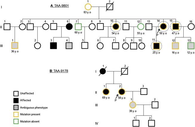
Thoracic aortic aneurysm and dissection (TAAD) is an autosomal-dominant disorder with major life-threatening complications. The disease displays great genetic heterogeneity with some forms allelic to Marfan and Loeys-Dietz syndrome, and an important number of cases still remain unexplained at the molecular level. Through whole-exome sequencing of affected members in a large TAAD-affected family, we identified the c.472C>T (p.Arg158(∗)) nonsense mutation in MFAP5 encoding the extracellular matrix component MAGP-2. This protein interacts with elastin fibers and the microfibrillar network. Mutation screening of 403 additional probands identified an additional missense mutation of MFAP5 (c.62G>T [p.Trp21Leu]) segregating with the disease in a second family. Functional analyses performed on both affected individual's cells and in vitro models showed that these two mutations caused pure or partial haploinsufficiency. Thus, alteration of MAGP-2, a component of microfibrils and elastic fibers, appears as an initiating mechanism of inherited TAAD.


Figures

Figure 1
Figure 1
Pedigrees of Families TAA-9801 and TAA-9178 Black arrows indicate probands. DNA from circled individuals was used for whole-exome sequencing. Orange symbols indicate family members heterozygous for mutations in MFAP5: c.472C>T (p.Arg158) in TAA-9801 (A) and c.62G>T (p.Trp21Leu) in TAA-9178 (B). The age at diagnosis is specified when available.
Figure 2
Figure 2
Structure of MFAP5 and MAGP-2 MFAP5 consists of ten exons (squares) with nine coding exons (blue squares). Mutations detected in this study appear under the gene schematic. MFAP5 encodes MAGP-2 for which the protein structure is depicted above the genomic structure. Scissors indicate the putative cleavage sites for the signal peptide (SP) peptidase and for proprotein convertases (RLRR). Putative site for cell attachment (RGD) and N- and O-Glycosylations (N-Glyc and O-Glyc) are also indicated. The electrophoregrams of each mutation obtained from Sanger sequencing appear below the genomic structure. The affected codons are underlined in black. Substitution c.62G>T (left) affects an evolutionary conserved tryptophan among mouse, rat, dog, cat, and chicken. Multiple sequence alignment was performed with CLUSTAL Omega. Comparative sequencing of mutation c.472C>T from genomic DNA and transcript of MFAP5 (cDNA) are presented (right). cDNA from individual TAA-9801 II:15 was obtained from mRNA harvested from primary fibroblasts grown with or without emetine to block the NMD RNA surveillance pathway.
Figure 3
Figure 3
Immunoblot Analysis of MAGP-2 in Fibroblasts from Affected Individuals Proteins were extracted from cellular lysates (A) and extracellular media (B) of fibroblasts from affected subjects carrying the nonsense (TAA-9801 family) or missense (TAA-9178) mutations and healthy control subjects. Intracellular soluble proteins were extracted with TRIzol reagent (Life Technologies) according to the manufacturer’s instructions. This method avoids contamination by insoluble cell-associated matrix such as matrix-associated MAGP-2. Extracellular proteins were extracted from extracellular media after centrifugation with the AMICON 10 kDa filtration columns (Millipore). Immunoblots were realized via two antibodies recognizing either N- or C-terminal domains of MAGP-2 as mutations affect N-terminal (p.Trp21Leu) or C-terminal (p.Arg158) domains or horseradish peroxidase-conjugated β-tubulin (1:5,000 dilution, ab21058 from Abcam). Purified recombinant human MAGP-2 expressed in HEK cells (TP303242 from Origene Technologies) was used as control on each blot. Densitometric analysis was performed with Scion Image software (Scion Corporation). Immunoblots showed the presence in cellular lysates of both WT and truncated MAGP-2 in individuals heterozygous for the nonsense c.472C>T variant. (C) Quantification of immunoblots (N-term) showed that the band corresponding to WT MAGP-2 represents less than 50% of the total MAGP-2 signal detected (normalized with β-tubulin; means ± standard error of the mean). The band corresponding to the truncated protein was not found in extracellular medium.
Figure 4
Figure 4
Immunoblot Analysis of WT, p.Arg158, and p.Trp21Leu MAGP-2 in Transfected HEK (A and B) Cellular lysates (A) and extracellular media (B) of transfected HEK with WT or c.472C>T (p.Arg158) coding vectors were analyzed. Transient transfection assays of constructs were performed in HEK293 H cells (HEK, GIBCO Life Technologies) because they do not express endogenous MFAP5. Cells were transiently cotransfected in serum-free DMEM (GIBCO Life Technologies) with 1 μg of a vector encoding WT or mutated c.472C>T (p.Arg158) MAGP-2 and 1 μg of an empty vector (pcDNA3.1). The Fugene 6 transfection agent was used (Promega). After transfection, HEK cells were incubated for 24 hr. The extracellular medium was harvested and cells were washed three times with PBS prior to RNA/protein extractions. Transfection efficiency was checked and showed proper expression of the MFAP5 transcripts for WT and mutant constructs (Figure S1). Protein extraction and IBs were performed as described in Figure 3. WT MAGP-2 was detected in both cellular lysates and extracellular media as expected. p.Arg158 truncated MAGP-2 protein was detectable in cellular lysates and totally absent from the extracellular medium. (C and D) Cellular lysates (C) and extracellular media (D) of transfected HEK with WT or c.62G>T (p.Trp21Leu) coding vectors were analyzed. Immunoblots and checking of transfection efficacy were realized as previously described. Immunoblotting of the N-terminal part of MAGP-2 was not performed because the mutation affects an amino acid located in the epitope. (E) Quantification of immunoblots confirmed a significant decrease of intracellular presence of p.Trp21Leu MAGP-2. All values are expressed as means ± standard error of the mean. Statistical differences between WT and p.Trp21Leu MAGP-2 were analyzed by a Student’s t test. The absence of p.Arg158 MAGP-2 precluded statistical analysis of WT versus p.Arg158. Differences were considered statistically significant at values of p < 0.05. Data shown for each construct were pooled from three independent transfection assays.
Figure 5
Figure 5
Immunohistology of Aortic Wall from a Dissection Case in Family TAA-9178 Aneurysmal ascending aorta was collected during aortic surgery for the proband from the TAA-9178 family (Bichat University Hospital) (II:2, heterozygous for the c.62G>T MFAP5 mutation). Normal thoracic aorta was obtained from a normal organ transplant donor with the authorization of the French Biomedicine Agency. Aneurysmal tissue was sampled in the outer curvature, the most dilated part of the ascending aorta. Aortic specimens were fixed in Bouin’s solution, and serial sections (6 μm thickness) were obtained from each specimen. (A) Masson’s trichrome and Alcian blue revealed a disorganization of the tunica media with a loss of smooth muscle cells (SMCs) and proteoglycan accumulation, respectively, compared to healthy aorta. (B) Immunohistofluorescent staining for the same subject and control samples was performed with antibodies to nuclear phosphorylated SMAD2/3 (phosphorylated Ser 423/425 SMAD2/3, sc-11769 from Santa Cruz Biotechnology) and mouse monoclonal antibody to TGF-β1 (sc-65378 from Santa Cruz Biotechnology). Alexa Fluor 555-conjugated secondary antibodies to mouse and rabbit (Invitrogen, Life Technologies) were used. Because pathology samples had been fixed in Bouin’s solution, SMC nuclei could not be Dapi-stained. Elastic fibers appear in green (TGF-β1) and yellow (p-SMAD2/3). Staining showed enhanced TGF-β signaling in patient compared to healthy aorta.

References

    1. Milewicz D.M., Guo D.C., Tran-Fadulu V., Lafont A.L., Papke C.L., Inamoto S., Kwartler C.S., Pannu H. Genetic basis of thoracic aortic aneurysms and dissections: focus on smooth muscle cell contractile dysfunction. Annu. Rev. Genomics Hum. Genet. 2008;9:283–302. - PubMed
    1. Jondeau G., Boileau C. Genetics of thoracic aortic aneurysms. Curr. Atheroscler. Rep. 2012;14:219–226. - PubMed
    1. Faivre L., Collod-Beroud G., Loeys B.L., Child A., Binquet C., Gautier E., Callewaert B., Arbustini E., Mayer K., Arslan-Kirchner M. Effect of mutation type and location on clinical outcome in 1,013 probands with Marfan syndrome or related phenotypes and FBN1 mutations: an international study. Am. J. Hum. Genet. 2007;81:454–466. - PMC - PubMed
    1. Mizuguchi T., Collod-Beroud G., Akiyama T., Abifadel M., Harada N., Morisaki T., Allard D., Varret M., Claustres M., Morisaki H. Heterozygous TGFBR2 mutations in Marfan syndrome. Nat. Genet. 2004;36:855–860. - PMC - PubMed
    1. Loeys B.L., Schwarze U., Holm T., Callewaert B.L., Thomas G.H., Pannu H., De Backer J.F., Oswald G.L., Symoens S., Manouvrier S. Aneurysm syndromes caused by mutations in the TGF-beta receptor. N. Engl. J. Med. 2006;355:788–798. - PubMed

Publication types

MeSH terms

Supplementary concepts

LinkOut - more resources