. 2015 Mar;407(8):2127-39.
doi: 10.1007/s00216-014-8293-7. Epub 2014 Dec 2.

MALDI imaging mass spectrometry of N-linked glycans on formalin-fixed paraffin-embedded murine kidney

Affiliations

MALDI imaging mass spectrometry of N-linked glycans on formalin-fixed paraffin-embedded murine kidney

Ove J R Gustafsson et al. Anal Bioanal Chem. 2015 Mar.

Abstract

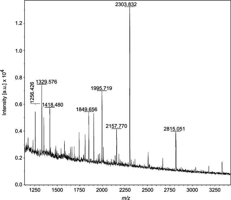
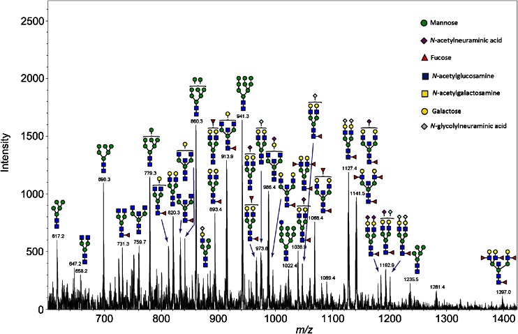
Recent developments in spatial proteomics have paved the way for retrospective in situ mass spectrometry (MS) analyses of formalin-fixed paraffin-embedded clinical tissue samples. This type of analysis is commonly referred to as matrix-assisted laser desorption/ionization (MALDI) imaging. Recently, formalin-fixed paraffin-embedded MALDI imaging analyses were augmented to allow in situ analyses of tissue-specific N-glycosylation profiles. In the present study, we outline an improved automated sample preparation method for N-glycan MALDI imaging, which uses in situ PNGase F-mediated release and measurement of N-linked glycans from sections of formalin-fixed murine kidney. The sum of the presented data indicated that N-glycans can be cleaved from proteins within formalin-fixed tissue and characterized using three strategies: (i) extraction and composition analysis through on-target MALDI MS and liquid chromatography coupled to electrospray ionization ion trap MS; (ii) MALDI profiling, where N-glycans are released and measured from large droplet arrays in situ; and (iii) MALDI imaging, which maps the tissue specificity of N-glycans at a higher resolution. Thus, we present a complete, straightforward method that combines MALDI imaging and characterization of tissue-specific N-glycans and complements existing strategies.


Figures

Graphical Abstract
Graphical Abstract
MALDI imaging MS of N-linked glycans released from formalin-fixed paraffin-embedded murine kidney sections. Ion intensity maps for (Hex)2(HexNAc)3(Deoxyhexose)3+(Man)3(GlcNAc)2 (m/z 2304.932, red), (Hex)6+(Man)3(GlcNAc)2 (m/z 1905.742, green) and (Hex)2(HexNAc)2+(Man)3(GlcNAc)2 (m/z 1663.756, blue)
Fig. 1
Fig. 1
In situ MS profiling of N-linked glycans released from formalin-fixed murine kidney sections using dialyzed PNGase F. Four replicate formalin-fixed murine kidney tissue sections were treated with antigen retrieval followed by ChIP-1000 printing of PNGase F or buffer control. An N-glycan calibrant mixture was spotted onto a separate part of the section. Tissue was overlaid with 2,5-DHB in 0.1 % trifluoroacetic acid and 1 mM NaCl using a TM sprayer instrument and MS spectra were acquired using a MALDI-TOF/TOF. a Hematoxylin and eosin (H & E)-stained murine kidney section annotated with the cortex, medulla, and pelvic regions. Cortex-medulla interface marked by a red dotted line (see Oetjen et al. [25]). b Overlay of H & E and profiling spots (blue dotted outline) treated with PNGase F (middle), buffer control (top), and calibrant spots (bottom). Ion intensity map in b is for mass filter 2304.778 (raw data loaded, baseline subtracted, data reduced to 20,000 points per spectrum, RMS normalization). The intensity map shows definitive signal in PNGase F-treated spots, with tissue-specific differences clear between spots localized to the cortex and those in the medulla. c, d The 4× control and 4× PNGase F MS spectra, respectively, for this section. Spectra were exported (flexAnalysis) and formatted in MS PowerPoint and InkScape (www.inkscape.org). MS spectra show clear differences in peak number, with the PNGase F spots containing multiple unique masses. Numbers with asterisks indicate N-glycan candidates identified by LC-MS/MS—these are also annotated in Table 2
Fig. 2
Fig. 2
MALDI-TOF/TOF MS and MS/MS of N-glycans released in-solution from formalin-fixed murine kidney sections. MS profile obtained from N-glycans enzymatically released (PNGase F) in-solution from FFPE kidney tissue treated with antigen retrieval. Spectrum was acquired on a MALDI-TOF/TOF MS instrument in reflectron positive ion mode and peaks were picked (no further processing). Formatting in MS PowerPoint and InkScape (www.inkscape.org). Multiple unique masses can be visualized in the PNGase F-treated tissue supernatant. (see corresponding m/z of potential N-linked glycans in Table 2)
Fig. 3
Fig. 3
Ion chromatogram annotated with structure proposals for N-glycans identified by ESI-ion trap MS/MS. N-glycans released in-solution from formalin-fixed murine kidney tissue using PNGase F were reduced, desalted, and purified on a carbon column prior to porous graphitic column chromatography coupled online to an ESI-ion trap (IT) system. MS scan above is annotated with the structure proposals that resulted from MS/MS structure profiling of the eluate
Fig. 4
Fig. 4
MALDI imaging MS of N-linked glycans released from formalin-fixed murine kidney sections. Formalin-fixed murine kidney sections were treated with citric acid antigen retrieval prior to printing of 30 nL/spot dialyzed PNGase F or buffer control arrays with 250 μm spacing onto the tissue. 2,5-DHB (20 mg/mL) was sprayed onto the tissue sections, and MS spectra were acquired by oversampling at 100 μm intervals using a MALDI-TOF/TOF MS instrument. Hematoxylin and eosin stained tissue sections are shown for PNGase F-treated (a, b) and buffer control-treated (c) regions, respectively. df DIPPS maps of the 13 m/z values with the highest DIPPS score (i.e., highest proportion in PNGase F-treated regions as compared to control) for the PNGase F (d, e) and buffer control (f) regions, respectively. gi The log ion intensity maps for one of these 13 m/z values, in this case 2304.909, for the PNGase F (g, h) and buffer control (i) regions. Both DIPPS maps and log ion intensity maps show increased counts/intensity in the cortex of the kidney
Fig. 5
Fig. 5
MALDI imaging MS of N-linked glycans released from formalin-fixed murine kidney sections. Formalin-fixed murine kidney sections were treated with antigen retrieval prior to printing of 30 nL/spot dialyzed PNGase F or buffer control arrays with 250 μm spacing. 2,5-DHB (20 mg/mL) was sprayed onto the sections and MS spectra were acquired by oversampling at 100 μm intervals using a MALDI-TOF/TOF MS instrument. Data was loaded raw into SCiLS, preprocessed for baseline subtraction and normalization to total ion current (TIC) prior to segmentation analysis (maximum processing mode, interval width of 0.5 Da, strong smoothing) and pLSA (10 component, interval width of 0.5 Da, random initiation). Regions are outlined for PNGase F regions 1 (red) and 2 (blue), as well as control (green) and calibrant (small red box) regions. a The segmentation map for the entire data set, which discriminates between the cortex and medulla/pelvis of the kidney at the segmentation levels selected (see inset). bd Components 1, 2, and 8 from the pLSA analysis, which also discriminate the same regions of the tissue. eh Ion intensity maps for (Hex)2 (HexNAc)2 + (Man)3(GlcNAc)2 (m/z 1663.632), (Hex)6 + (Man)3(GlcNAc)2 (m/z 1905.697), (Hex)2(HexNAc)3(Deoxyhexose)3 + (Man)3(GlcNAc)2 (m/z 2304.909), and (Hex)3(HexNAc)4 (Deoxyhexose)4 + (Man)3(GlcNAc)2 (m/z 2816.115). Intensity scales are autocorrected for these intensity maps (with weak denoising) and scale bars are included

References

    1. Pan S, Chen R, Aebersold R, Brentnall TA (2011) Mass spectrometry based glycoproteomics—from a proteomics perspective. Molecular & Cellular Proteomics: MCP 10 (1):R110 003251. doi:10.1074/mcp.R110.003251 - PMC - PubMed
    1. Kam RKT, Poon TCW. The potentials of glycomics in biomarker discovery. Clin Proteom. 2008;4:67–79. doi: 10.1007/s12014-008-9017-9. - DOI
    1. Kolarich D, Lepenies B, Seeberger PH. Glycomics, glycoproteomics and the immune system. Curr Opin Chem Biol. 2012;16(1–2):214–220. doi: 10.1016/j.cbpa.2011.12.006. - DOI - PubMed
    1. Abbott KL, Nairn AV, Hall EM, Horton MB, McDonald JF, Moremen KW, Dinulescu DM, Pierce M. Focused glycomic analysis of the N-linked glycan biosynthetic pathway in ovarian cancer. Proteomics. 2008;8(16):3210–3220. doi: 10.1002/pmic.200800157. - DOI - PMC - PubMed
    1. Crockett DK, Lin Z, Vaughn CP, Lim MS, Elenitoba-Johnson KS. Identification of proteins from formalin-fixed paraffin-embedded cells by LC-MS/MS. Lab Invest. 2005;85(11):1405–1415. doi: 10.1038/labinvest.3700343. - DOI - PubMed

Publication types