. 2015 Jan;36(1):131-8.
doi: 10.1038/aps.2014.108. Epub 2014 Dec 1.

β, β-Dimethylacrylshikonin induces mitochondria-dependent apoptosis of human lung adenocarcinoma cells in vitro via p38 pathway activation

Affiliations

β, β-Dimethylacrylshikonin induces mitochondria-dependent apoptosis of human lung adenocarcinoma cells in vitro via p38 pathway activation

Hai-bing Wang et al. Acta Pharmacol Sin. 2015 Jan.

Abstract

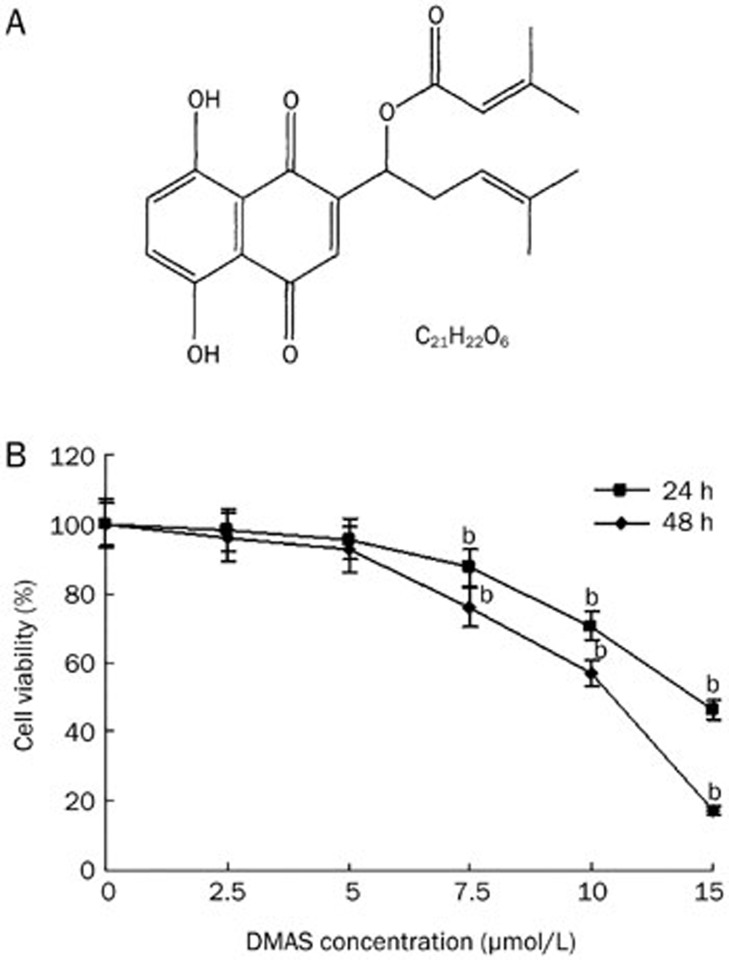
Aim: β, β-Dimethylacrylshikonin (DMAS) is an anticancer compound extracted from the roots of Lithospermum erythrorhizon. In the present study, we investigated the effects of DMAS on human lung adenocarcinoma cells in vitro and explored the mechanisms of its anti-cancer action.

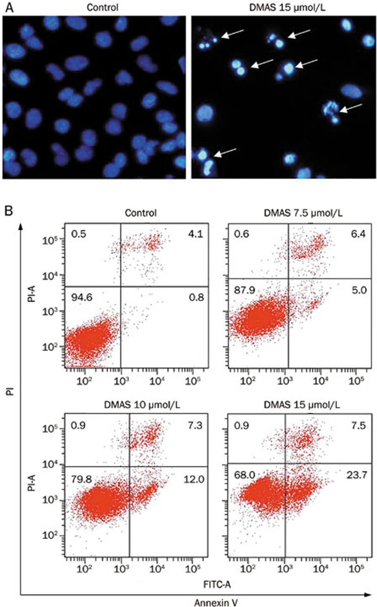
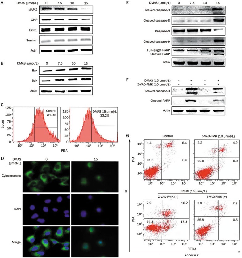
Methods: Human lung adenocarcinoma A549 cells were tested. Cell viability was assessed using an MTT assay, and cell apoptosis was evaluated with flow cytometry and DAPI staining. The expression of the related proteins was detected using Western blotting. The mitochondrial membrane potential was measured using a JC-1 kit, and subcellular distribution of cytochrome c was analyzed using immunofluorescence staining.

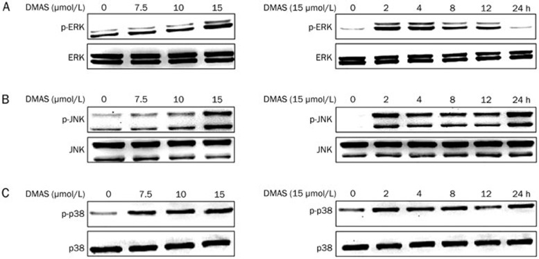
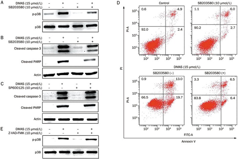
Results: Treatment of A549 cells with DMAS suppressed the cell viability in dose- and time-dependent manners (the IC50 value was 14.22 and 10.61 μmol/L, respectively, at 24 and 48 h). DMAS (7.5, 10, and 15 μmol/L) dose-dependently induced apoptosis, down-regulated cIAP-2 and XIAP expression, and up-regulated Bax and Bak expression in the cells. Furthermore, DMAS resulted in loss of mitochondrial membrane potential and release of cytochrome c in the cells, and activated caspase-9, caspase-8, and caspase-3, and subsequently cleaved PARP, which was abolished by pretreatment with Z-VAD-FMK, a pan-caspase inhibitor. DMAS induced sustained p38 phosphorylation in the cells, while pretreatment with SB203580, a specific p38 inhibitor, blocked DMAS-induced p38 activation and apoptosis.

Conclusion: DMAS inhibits the growth of human lung adenocarcinoma A549 cells in vitro via activation of p38 signaling pathway.


Figures

Figure 1
Figure 1
DMAS inhibits A549 cell growth. (A) The chemical structure of DMAS (MW370.4). (B) Effects of DMAS on the inhibition of A549 cell viability. The cells were treated with DMAS at different concentrations for 24 and 48 h. The cell viability was determined using an MTT assay. The viability of the control group (0.1% DMSO) was set to 100%. The data represent the mean±SD obtained from three independent experiments. bP<0.05 compared with the control group.
Figure 2
Figure 2
DMAS induced apoptosis in A549 cells. (A) DNA condensation (white arrow) was measured through DAPI staining and analyzed under a fluorescent microscope at 200× magnification. (B) The apoptotic status was evaluated using an Annexin V-FITC binding assay. Early stage apoptotic cells are shown on the lower right (Annexin V-FITC+/PI), and late stage apoptotic cells are shown on the top right. The part (Annexin V-FITC/PI) was considered as Viable cells are shown on the lower left, and necrotic cells are shown on the upper left (Annexin V-FITC/PI+).
Figure 3
Figure 3
DMAS induced mitochondrial events associated with apoptosis in A549 cells. Western blot analysis for anti-apoptotic proteins: Bcl-xL, cIAP-2, XIAP, and survivin (A) and pro-apoptotic proteins: Bax and Bak (B) in whole cell extracts of A549 cells treated with DMAS for 24 h. Actin protein levels were also measured as controls. (C) The detection of mitochondrial membrane potential through flow cytometry. A549 cells were treated with or without DMAS (15 μmol/L) for 24 h, followed by staining with JC-1 for 15 min at 37 °C and flow cytometry. (D) A549 cells were fixed and labeled for cytochrome c (green) and DNA (blue). (E) Western blot analysis for cleaved PARP, cleaved caspase-9, cleaved caspase-8, cleaved caspase-3 in whole cell extracts of A549 cells treated with DMAS for 24 h. Actin protein levels were also measured as controls. (F) A549 cells were treated with DMAS in the presence or absence of Z-VAD-FMK (10 μmol/L) for 24 h. The protein extracts were prepared and subjected to Western blot analysis using antibodies against cleaved caspase-3 and cleaved PARP. Actin protein levels were also measured as controls. (G) A549 cells were treated with DMAS in the presence or absence of Z-VAD-FMK (10 μmol/L) for 24 h. The apoptotic status was determined using an Annexin V-FITC binding assay.
Figure 4
Figure 4
Effects of DMAS on the MAPK signaling pathway. A549 cells were treated with DMAS at the indicated concentrations for the indicated times, and total cellular extracts were prepared and subjected to Western blot analysis to measure the levels of phosphorylated ERK (A), JNK (B), and p38 (C). The membranes were reprobed with antibodies against total ERK, JNK, and p38 for normalization.
Figure 5
Figure 5
DMAS-induced A549 cell apoptosis is mediated through p38 activation. (A) A549 cells were pretreated with or without SB203580 (10 μmol/L), followed by treatment with DMSO (vehicle) or DMAS (15 μmol/L) for 24 h. Protein extracts were prepared and subjected to Western blot analysis to measure the levels of phosphorylated p38. Total p38 protein levels were also measured as controls. (B) A549 cells were pretreated or not with SB203580 (10 μmol/L), followed by treatment with DMSO (vehicle) or DMAS (15 μmol/L) for 24 h. Protein extracts were prepared and subjected to Western blot analysis using antibodies against cleaved caspase-3 and cleaved PARP. Actin protein levels were also measured as controls. (C) A549 cells were pretreated or not with SP600125 (25 μmol/L), followed by treatment with DMSO (vehicle) or DMAS (15 μmol/L) for 24 h. Protein extracts were prepared and subjected to Western blot analysis using antibodies against cleaved caspase-3 and cleaved PARP. Actin protein levels were also measured as controls. (D) A549 cells were treated with DMAS in the presence or absence of SB203580 (10 μmol/L) for 24 h. The apoptotic status was determined using an Annexin V-FITC binding assay. (E) A549 cells were pretreated with or without Z-VAD-FMK (10 μmol/L), followed by treatment with DMAS (15 μmol/L) for 24 h. Protein extracts were prepared and subjected to Western blot analysis to measure the levels of phosphorylated p38. Total p38 protein levels were also measure as controls.

References

    1. Hanagiri T, Baba T, So T, Yasuda M, Sugaya M, Ono K, et al. Time trends of surgical outcome of patients with non-small cell lung cancer. J Thorac Oncol 2010; 5: 825–9. - PubMed
    1. Xuan Y, Hu X. Naturally-occurring shikonin analogues — a class of necroptotic inducers that circumvent cancer drug resistance. Cancer Lett 2009; 274: 233–42. - PubMed
    1. Shen CC, Syu WJ, Li SY, Lin CH, Lee GH, Sun CM. Antimicrobial activities of naphthazarins from Arnebia euchroma. J Nat Prod 2002; 65: 1857–62. - PubMed
    1. Tanaka S, Tajima M, Tsukada M, Tabata M. A comparative study on anti-inflammatory activities of the enantiomers, shikonin and alkannin. J Nat Prod 1986; 49: 466–9. - PubMed
    1. Chen X, Yang L, Zhang N, Turpin JA, Buckheit RW, Osterling C, et al. Shikonin, a component of Chinese herbal medicine, inhibits chemokine receptor function and suppresses human immunodeficiency virus type 1. Antimicrob Agents Chemother 2003; 47: 2810–6. - PMC - PubMed

Publication types

MeSH terms

Substances