Natural killer T cells play a necessary role in modulating of immune-mediated liver injury by gut microbiota
- PMID: 25435303
- PMCID: PMC4248284
- DOI: 10.1038/srep07259
Natural killer T cells play a necessary role in modulating of immune-mediated liver injury by gut microbiota
Abstract
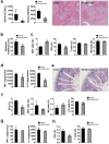
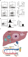
Gut microbiota are implicated in many liver diseases. Concanavalin A (ConA)-induced hepatitis is a well-characterized murine model of fulminant immunological hepatic injury. Oral administration of pathogenic bacteria or gentamycin to the mice before ConA injection, liver injury and lymphocyte distribution in liver and intestine were assessed. Our data show that administration of pathogenic bacteria exacerbated the liver damage. There was more downregulation of activation-induced natural killer T (NKT) cells in the liver of pathogenic bacteria-treated ConA groups. Also, there was a negative correlation between the numbers of hepatic NKT cells and liver injury in our experiments. Moreover, intestinal dendritic cells (DCs) were increased in pathogenic bacteria-treated ConA groups. The activation of DCs in Peyer's patches and the liver was similar to the intestine. However, depletion of gut gram-negative bacteria alleviated ConA-induced liver injury, through suppressed hepatic NKT cells activation and DCs homing in liver and intestine. In vitro experiments revealed that DCs promoted NKT cell cytotoxicity against hepatocyte following stimulation with pathogenic bacteria. Our study suggests that increased intestinal pathogenic bacteria facilitate immune-mediated liver injury, which may be due to the activation of NKT cells that mediated by intestinal bacterial antigens activated DCs.
Figures


Similar articles
-
Critical roles of conventional dendritic cells in promoting T cell-dependent hepatitis through regulating natural killer T cells.Clin Exp Immunol. 2017 Apr;188(1):127-137. doi: 10.1111/cei.12907. Epub 2017 Jan 5. Clin Exp Immunol. 2017. PMID: 27891589 Free PMC article.
-
PARP2 deficiency affects invariant-NKT-cell maturation and protects mice from concanavalin A-induced liver injury.Am J Physiol Gastrointest Liver Physiol. 2017 Nov 1;313(5):G399-G409. doi: 10.1152/ajpgi.00436.2016. Epub 2017 Jul 27. Am J Physiol Gastrointest Liver Physiol. 2017. PMID: 28751426
-
Enterogenous bacterial glycolipids are required for the generation of natural killer T cells mediated liver injury.Sci Rep. 2016 Nov 8;6:36365. doi: 10.1038/srep36365. Sci Rep. 2016. PMID: 27821872 Free PMC article.
-
Natural killer T cells within the liver: conductors of the hepatic immune orchestra.Dig Dis. 2010;28(1):7-13. doi: 10.1159/000282059. Epub 2010 May 7. Dig Dis. 2010. PMID: 20460885 Review.
-
Complex Network of NKT Cell Subsets Controls Immune Homeostasis in Liver and Gut.Front Immunol. 2018 Sep 11;9:2082. doi: 10.3389/fimmu.2018.02082. eCollection 2018. Front Immunol. 2018. PMID: 30254647 Free PMC article. Review.
Cited by
-
Gut microbiota modulate the immune effect against hepatitis B virus infection.Eur J Clin Microbiol Infect Dis. 2015 Nov;34(11):2139-47. doi: 10.1007/s10096-015-2464-0. Epub 2015 Aug 14. Eur J Clin Microbiol Infect Dis. 2015. PMID: 26272175 Review.
-
The Role of Gut Microbiota in Some Liver Diseases: From an Immunological Perspective.Front Immunol. 2022 Jul 13;13:923599. doi: 10.3389/fimmu.2022.923599. eCollection 2022. Front Immunol. 2022. PMID: 35911738 Free PMC article. Review.
-
NK1.1+ cells promote sustained tissue injury and inflammation after trauma with hemorrhagic shock.J Leukoc Biol. 2017 Jul;102(1):127-134. doi: 10.1189/jlb.3A0716-333R. Epub 2017 May 17. J Leukoc Biol. 2017. PMID: 28515228 Free PMC article.
-
Cellular Innate Immunity: An Old Game with New Players.J Innate Immun. 2017;9(2):111-125. doi: 10.1159/000453397. Epub 2016 Dec 23. J Innate Immun. 2017. PMID: 28006777 Free PMC article. Review.
-
Natural Killer T Cells Contribute to Neutrophil Recruitment and Ocular Tissue Damage in a Model of Intraocular Tumor Rejection.Invest Ophthalmol Vis Sci. 2016 Mar;57(3):813-23. doi: 10.1167/iovs.15-18786. Invest Ophthalmol Vis Sci. 2016. PMID: 26934137 Free PMC article.
References
-
- Diao H. et al. Osteopontin as a mediator of NKT cell function in T cell-mediated liver diseases. Immunity 21, 539–550 (2004). - PubMed
-
- Miele L. et al. Increased intestinal permeability and tight junction alterations in nonalcoholic fatty liver disease. Hepatology 49, 1877–1887 (2009). - PubMed
-
- De Minicis S. et al. Dysbiosis contributes to fibrogenesis in the course of chronic liver injury in mice. Hepatology 59, 1738–1749 (2014). - PubMed
Publication types
MeSH terms
LinkOut - more resources
Full Text Sources
Other Literature Sources
Medical
Molecular Biology Databases
Research Materials

