Review
. 2013 May 13;2(2):288-356.
doi: 10.3390/pathogens2020288.

From in vitro to in vivo Models of Bacterial Biofilm-Related Infections

Affiliations
Review

From in vitro to in vivo Models of Bacterial Biofilm-Related Infections

David Lebeaux et al. Pathogens. .

Abstract

The influence of microorganisms growing as sessile communities in a large number of human infections has been extensively studied and recognized for 30-40 years, therefore warranting intense scientific and medical research. Nonetheless, mimicking the biofilm-life style of bacteria and biofilm-related infections has been an arduous task. Models used to study biofilms range from simple in vitro to complex in vivo models of tissues or device-related infections. These different models have progressively contributed to the current knowledge of biofilm physiology within the host context. While far from a complete understanding of the multiple elements controlling the dynamic interactions between the host and biofilms, we are nowadays witnessing the emergence of promising preventive or curative strategies to fight biofilm-related infections. This review undertakes a comprehensive analysis of the literature from a historic perspective commenting on the contribution of the different models and discussing future venues and new approaches that can be merged with more traditional techniques in order to model biofilm-infections and efficiently fight them.


Figures

Figure 1
Figure 1
Most studied biofilm-related infections in humans. Adapted from [17].
Figure 2
Figure 2
Non-mammalian in vivo models. A. Experimental settings. Drosophila melanogaster. Ten male fruit flies are selected and introduced in standard fly vials. A dilution of a Vibrio cholerae overnight culture to 5 * 108 CFU/mL is used to impregnate a 0.5-inch cellulose acetate plug placed at the bottom of each vial. Then, the vials are kept at 24 °C with appropriate light-dark cycles. Fruit fly survival is monitored twice a day for 5 to 7 days. B. Confocal microscopy image of D. melanogaster rectum papillae (oval structures) colonized by a V. cholerae (gfp-tagged, green) biofilm. Cell nuclei are stained in blue (DAPI staining). Images Credit: A. Purdy and P.I. Watnick Division of Infectious Diseases, Children’s Hospital, Boston, USA. Adapted from [38]. C. Experimental settings. Axenic zebrafish infection. After fertilization, eggs are immediately sterilized and kept in vented cap cell culture flasks in autoclaved mineral water at 28 °C. Starting at 4 dpf (days after fertilization), larvae are fed every 2 days with axenic Tetrahymena thermophila until day 15. For longer experiments, in addition to T. thermophila, larvae were fed axenic Artemia salina from 10 dpf onwards. Zebrafish larvae are infected 6 days after fertilization with 5 * 108 CFU/mL of pathogen. Mortality can be easily followed on daily basis. Adapted from [41]. D. Confocal fluorescence pictures of larval intestine infected by the pathogen E. ictaluri (detected by immunofluorescence, red) 1 day after infection. Zebrafish cell nuclei are shown in blue (DAPI staining) and actin in green. Images Credit: J.P. Levraud and M. Frétaud, Institut Pasteur, Paris, France.
Figure 3
Figure 3
Burn wound infection biofilm in mice model. A. Experimental Settings. Mice are subcutaneously anaesthetized, shaved and then covered with a fire blanket and a metal plate with a window corresponding to approximately 6% of total body surface. A third-degree burn is then induced using a hot-air blower for 7 s at 330 °C. Afterwards, mice receive fluid replacement and pain therapy during the whole experiment. Lastly, mice are infected by alginate embedded Pseudomonas aeruginosa beneath the burn wound 2–4 days after burn wound infliction. B. Clinical result 4 days after the procedure. Thermal third degree lesion associated with a wound infection. C. Confocal laser scanning microscopy of burn wound. A slide of the wound removed in toto is stained with P. aeruginosa specific peptide nucleic acid (PNA) fluorescence in situ hybridization (FISH) probe (magnification × 400). P. aeruginosa forms dense bacterial clusters (black arrowhead) on the surface of the burn wound. White arrowhead indicates subcutaneous area. Images Credit: C. Moser, K. Thomsen, H. Calum and H. Trøstrup, Department of Clinical Microbiology, Rigshospitalet, Denmark. Adapted from [154,155].
Figure 4
Figure 4
Native valve endocarditis in rabbit model. A. Post-mortem examination of a rabbit heart. Aortic endocarditis is induced in female New Zealand White rabbits by insertion of a polyethylene catheter (black arrow) through the right carotid artery into the left ventricle. Twenty-four hours after catheter insertion, pathogenic bacteria were inoculated through ear vein in each rabbit. The catheter is left in place throughout the experiment. Animals are killed 8 h after the last antibiotic injection and the vegetations (white arrowheads) from each rabbit are excised, rinsed in saline, pooled, and weighed. White arrow: left ventricle wall; black arrowhead: aorta; black star: aortic valve. B. Scanning electron microscopy of vegetation after 11 days of infection. Biofilm formed by Streptococcus spp. at the surface of native aortic valve. C. Transmission electron microscopy of bacteria from vegetation after 11 days of infection. Ruthenium red staining reveals the presence of an extracellular matrix (black arrowhead) surrounding Streptococcus spp. (white arrowhead) causing native aortic endocarditis. Images credit: A.-C. Crémieux (EA3647, Université Versailles Saint-Quentin), V. Dubée and B. Fantin (EA3964, Université Paris Diderot, Faculté de Médecine, Paris, France). Adapted from [167,168].
Figure 5
Figure 5
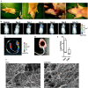
Totally implantable venous access port (TIVAP)-associated biofilm using rat model. A. Experimental Settings. Rats are anesthetized and shaved before starting the procedure. After skin disinfection, an incision is made at the dorsal midline, a subcutaneous pocket is created and the port is carefully inserted before being held intact by sutures. An incision is made in the neck area on the ventral side in order to access the external jugular vein. The catheter is inserted into the vein and pushed up to the superior vena cava. Suturing of both the dorsal and ventral sides closed the wounds and rats received analgesia at the end of the experiment. B. Monitoring of TIVAP colonization by E. coli. Five days after TIVAP insertion, 104 CFU of E. coli in 100 µL are injected into the port and photon emission is measured over a period of 10 days to monitor biofilm growth. Dorsal view of a representative rat, showing progression of biofilm signals towards the catheter tip and then restriction to the port. C, D, E and F. Bacterial colonization of TIVAP leads to biofilm formation. Rats are sacrificed 10 days post-infection and TIVAP are removed aseptically for examination. C. Photon emission of the removed TIVAP colonized by E. coli biofilm. D. Macroscopic examination after septum removal showing blood clots and deposits inside the port. E. Bacterial cells are harvested from the catheter and port separately and plated on LB agar for CFU counting. F. SEM images confirming biofilm formation in TIVAP in vivo in the port and catheter. Adapted from [256].
Figure 6
Figure 6
Model of endotracheal tube biofilm–associated infections in ventilated pigs. A. Experimental Settings. Large-White Landrace female pig (36 Kg) orotracheally intubated and mechanically ventilated. Following intubation, the animal received an oropharyngeal challenge of Pseudomonas aeruginosa. During mechanical ventilation, endogenous oropharyngeal bacteria and Pseudomonas aeruginosa rapidly colonize the internal surface of the endotracheal tube. Bacteria within the endotracheal tube constitute a persistent source of pathogens, which may result in ventilator-associated tracheobronchitis and pneumonia. B. Endotracheal tube internal surface following 72 hours of mechanical ventilation. After extubation, the endotracheal tube external surface is cleaned with sterile gauzes and decontaminated by careful rinsing with 80% alcohol and saline solution and then longitudinally sliced open. Two 1 cm long sections and one 3 cm long section of the dependent half of endotracheal tube are dissected for confocal electron microscopy, scanning electron microscopy and quantitative microbiological studies, respectively. C. Scanning electron microscopy (magnification × 2000) of the internal surface of endotracheal tube (lateral view). Bacterial communities are adherent to the endotracheal tube surface and surrounded by the extracellular matrix. D. Confocal laser scanning microscopy of the internal surface of endotracheal tube (lateral view). The lumen of endotracheal tube is stained with BacLight Live/Dead stain (magnification × 250). Pseudomonas aeruginosa biofilm is adherent on the internal surface of endotracheal tube. Eukaryotic cells are also present within the biomass. Images Credit: G. Li Bassi and L. Fernandez-Barat, Pulmonary and Critical Care Unit, Division of Animal Experimentation, Hospital Clinic, Barcelona. Adapted from [294,299].

References

    1. Henrici A.T. Studies of freshwater bacteria: I. A direct microscopic technique. J. Bacteriol. 1933;25:277–287. - PMC - PubMed
    1. Costerton J.W., Geesey G.G., Cheng K.J. How bacteria stick. Sci. Am. 1978;238:86–95. - PubMed
    1. Geesey G.G., Richardson W.T., Yeomans H.G., Irvin R.T., Costerton J.W. Microscopic examination of natural sessile bacterial populations from an alpine stream. Can. J. Microbiol. 1977;23:1733–1736. - PubMed
    1. Al-Azemi A., Fielder M.D., Abuknesha R.A., Price R.G. Effects of chelating agent and environmental stresses on microbial biofilms: relevance to clinical microbiology. J. Appl. Microbiol. 2011;110:1307–1313. - PubMed
    1. Hoiby N., Bjarnsholt T., Givskov M., Molin S., Ciofu O. Antibiotic resistance of bacterial biofilms. Int. J. Antimicrob. Agents. 2010;35:322–332. - PubMed