. 2014 Nov 6;9(3):801-10.
doi: 10.1016/j.celrep.2014.10.006. Epub 2014 Oct 30.

Tissue-specific posttranslational modification allows functional targeting of thyrotropin

Affiliations

Tissue-specific posttranslational modification allows functional targeting of thyrotropin

Keisuke Ikegami et al. Cell Rep. .

Abstract

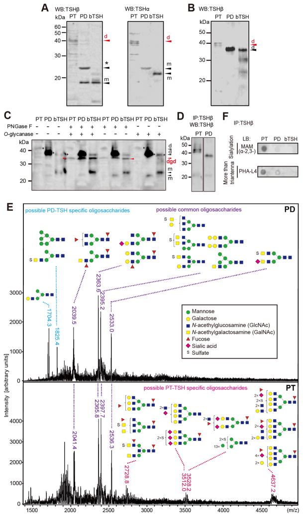
Thyroid-stimulating hormone (TSH; thyrotropin) is a glycoprotein secreted from the pituitary gland. Pars distalis-derived TSH (PD-TSH) stimulates the thyroid gland to produce thyroid hormones (THs), whereas pars tuberalis-derived TSH (PT-TSH) acts on the hypothalamus to regulate seasonal physiology and behavior. However, it had not been clear how these two TSHs avoid functional crosstalk. Here, we show that this regulation is mediated by tissue-specific glycosylation. Although PT-TSH is released into the circulation, it does not stimulate the thyroid gland. PD-TSH is known to have sulfated biantennary N-glycans, and sulfated TSH is rapidly metabolized in the liver. In contrast, PT-TSH has sialylated multibranched N-glycans; in the circulation, it forms the macro-TSH complex with immunoglobulin or albumin, resulting in the loss of its bioactivity. Glycosylation is fundamental to a wide range of biological processes. This report demonstrates its involvement in preventing functional crosstalk of signaling molecules in the body.


Figures

Figure 1
Figure 1. PT-TSH secreted into the peripheral circulation has little bioactivity
(A–D) Effects of melatonin on TSH concentration in PT (A), PD (B), and serum (C), and on serum T4 level (D). *P<0.05; **P<0.01 (t-test, n=6–12). (E–G) Effects of changing day length and T3 administration on Tshb mRNA (E), TSH concentration in PD and PT (F), and serum TSH and T4 levels (G). LD: long day; SD: short day; T3: daily T3 administration. *P<0.05; **P < 0.01; ##P<0.01 vs. smallest value (ANOVA, Fisher’s LSD post-hoc test, n=5–9). Values are means±s.e.m. Scale bars: 200 μm.
Figure 2
Figure 2. Glycosylation is different between PT-TSH and PD-TSH
(A) WB of PT and PD homogenates with TSHβ and TSHα antibodies under reducing and denaturing conditions. m: monomeric band; d: dimeric bands; *: non-specific bands. (B) WB of PT and PD homogenate under non-reducing conditions. (C) Deglycosylated TSHs were analyzed under non-reducing conditions. dgd: deglycosylated form; arrow: glycosylated form. (D) WB of IP-TSH under non-reducing conditions. (E) Positive-ion MALDI-TOF-MS of N-glycans cleaved from IP-TSHs. Blue: PD-TSH-specific N-glycans; Red: PT-TSH-specific N-glycans; Purple: common N-glycans. (F) LB analysis of TSHs.
Figure 3
Figure 3. TSH forms in the circulation macro-TSH with IgG or albumin
(A) WB of serum TSH from LD+T3 mice (See Figure 1). (B) Measurements of TSH, IgGs, and IgM in fractions from gel-filtration chromatography. Yellow shading: macro-TSH; arrow: additional peak. (C) Separation of IgG subclasses by Protein A affinity chromatography. Black open arrow: free TSH; black solid arrow: macro-TSH; red solid arrows: IgG2b; red open arrows: IgG2a, IgG3. (D) IgG isotyping of serum and macro-TSH. (E) Adsorption test of TSH with serum. *P<0.05; **P<0.01 vs. PD (means±s.e.m., t-test, n=4). (F,G) WB of TSH (F) and albumin (G) in fractions from Protein A affinity chromatography. Arrowhead: albumin-TSH complex. (H) Detection of IP-albumin-TSH complex in sera. Red: albumin-TSH complex; black: free albumin. (I) Macro-TSH disappeared when albumin was removed from the IgG-free flow-through fraction. Arrowhead: position of albumin-TSH. (J) WB of TSHβ and IgG2b in macro-TSH fractions under acidification condition (pH 3) by citrate or denature condition by DTT. Arrowhead: position of IgG-TSH.
Figure 4
Figure 4. Formation of macro-TSH strongly reduces bioactivity of PT-TSH
(A) Bioactivity of TSH from PT or PD homogenates, analyzed using TSHR-expressing CHO cells. Ratio of TSH bioactivity to immunoreactivity (B/I) was evaluated by cAMP accumulation. (B) Bioactivity of serum TSH from LD+T3 and SD animals, examined using primary cultured MBH and thyrocytes. (C) Bioactivity of macro-TSH (fr 20 of Figure 3B) and free TSH (fr 30), examined using primary cultured MBH and thyrocytes. (D) Effect of serum incubation on bioactivity of PT-TSH and PD-TSH. All values are means+s.e.m., n=3–5. *P<0.05; **P<0.01 (t-test).

Similar articles

Cited by

References

    1. Aksentijevich I, Sachs DH, Sykes M. Natural antibodies against bone marrow cells of a concordant xenogeneic species. J Immunol. 1991;147:4140–4146. - PubMed
    1. Andersen S, Pedersen KM, Bruun NH, Laurberg P. Narrow individual variations in serum T4 and T3 in normal subjects: a clue to the understanding of subclinical thyroid disease. J Clin Endocrinol Metab. 2002;87:1068–1072. - PubMed
    1. Baensiger JU, Green ED. Pituitary glycoprotein hormone oligosaccharides: structure, synthesis and function of the asparagine-linked oligosaccharides on lutropin, follitropin and thyrotropin. Biochim Biophys Acta. 1988;947:287–306. - PubMed
    1. Baker BL, Yu YY. Hypophyseal changes induced by thyroid deficiency and thyroxine administration as revealed by immunochemical staining. Endocrinology. 1971;89:996–1004. - PubMed
    1. Beck-Peccoz P, Amr S, Menezes-Ferreira MM, Faglia G, Weintraub BD. Decreased receptor binding of biologically inactive thyrotropin in central hypothyroidism. Effect of treatment with thyrotropin-releasing hormone. N Engl J Med. 1985;312:1085–1090. - PubMed

Publication types

LinkOut - more resources