. 2014 Nov 6;9(3):930-43.
doi: 10.1016/j.celrep.2014.10.008. Epub 2014 Oct 30.

Defining midbrain dopaminergic neuron diversity by single-cell gene expression profiling

Affiliations

Defining midbrain dopaminergic neuron diversity by single-cell gene expression profiling

Jean-Francois Poulin et al. Cell Rep. .

Abstract

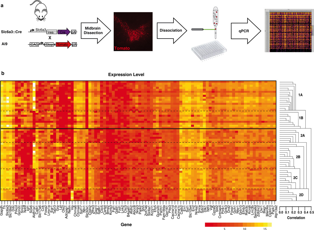
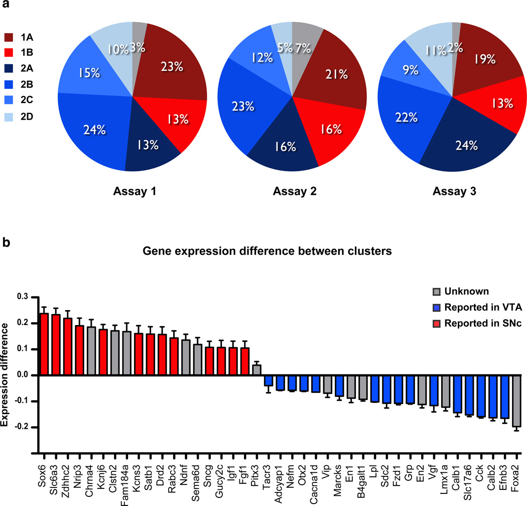
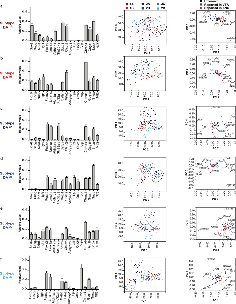
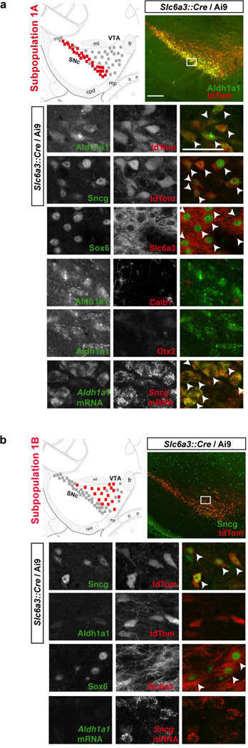
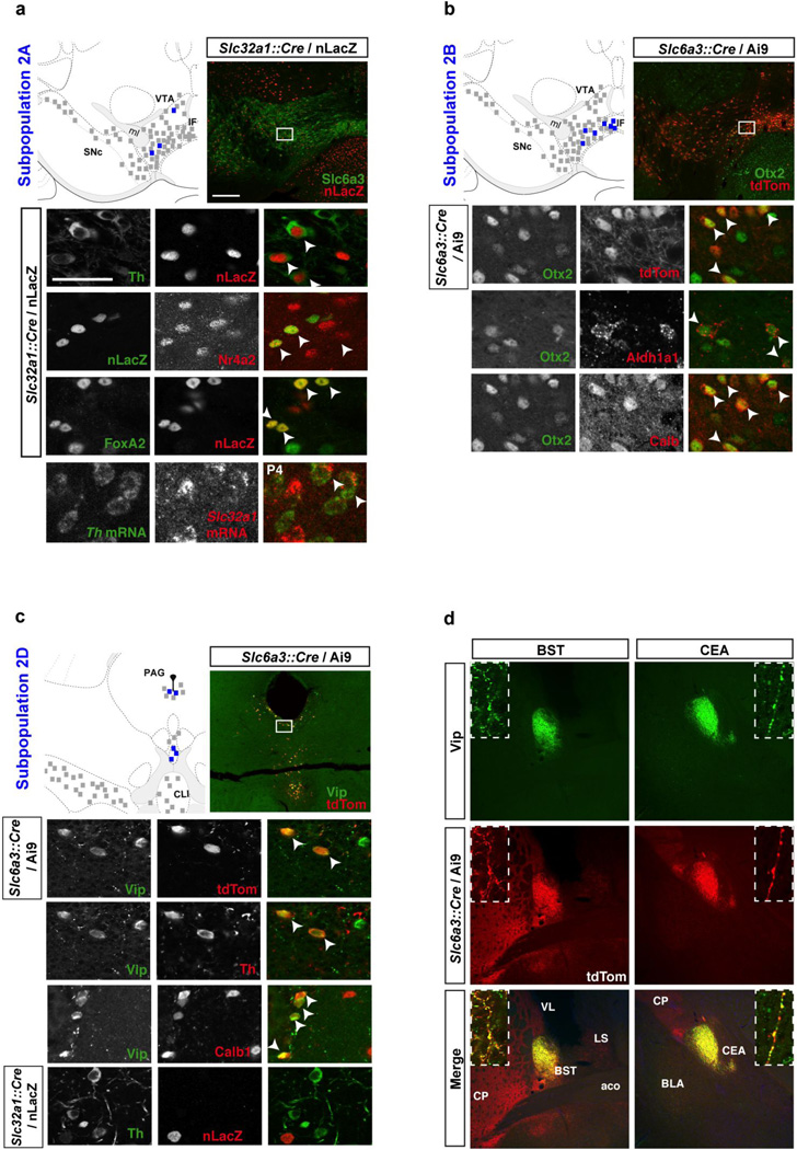
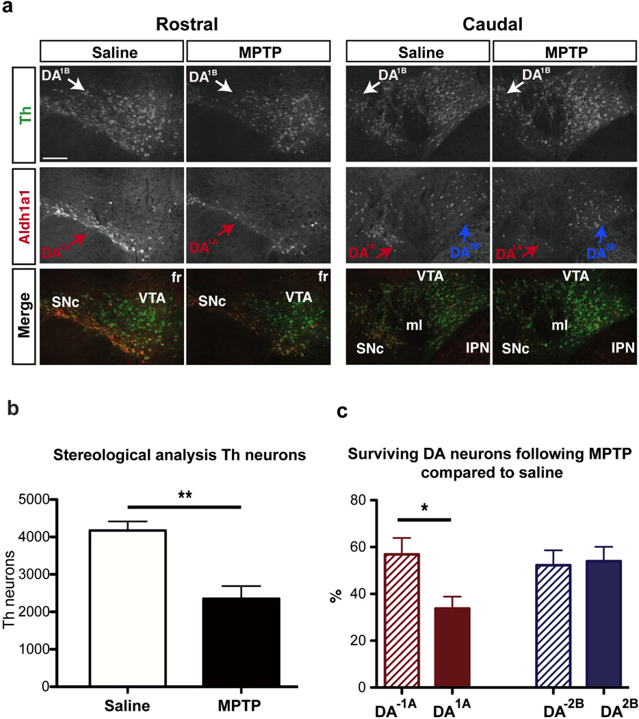
Effective approaches to neuropsychiatric disorders require detailed understanding of the cellular composition and circuitry of the complex mammalian brain. Here, we present a paradigm for deconstructing the diversity of neurons defined by a specific neurotransmitter using a microfluidic dynamic array to simultaneously evaluate the expression of 96 genes in single neurons. With this approach, we successfully identified multiple molecularly distinct dopamine neuron subtypes and localized them in the adult mouse brain. To validate the anatomical and functional correlates of molecular diversity, we provide evidence that one Vip+ subtype, located in the periaqueductal region, has a discrete projection field within the extended amygdala. Another Aldh1a1+ subtype, located in the substantia nigra, is especially vulnerable in the 1-methyl-4-phenyl-1,2,3,6-tetrahydropyridine (MPTP) model of Parkinson's disease. Overall, this rapid, cost-effective approach enables the identification and classification of multiple dopamine neuron subtypes, with distinct molecular, anatomical, and functional properties.


Figures

Figure 1
Figure 1. High throughput single-cell gene expression analysis of midbrain dopamine neurons
(a) The midbrain of postnatal day four Slc6a3::Cre; Ai9 midbrain was dissected and dissociated. Fluorescent (tdTomato+) single cells were FACS sorted into 96-well plate and analyzed for the expression of 96 genes using Fluidigm Biomark system. (b) A sample array in which the expression level of each gene is depicted on a logarithmic scale with red indicating low expression and yellow representing a high expression. Unbiased clustering analysis revealed the presence of two main clusters that could be further divided into a total of six molecularly distinct dopamine (DA) neuron subtypes.
Figure 2
Figure 2. Dopamine neurons cluster into distinct subtypes
(a) Out of 159 cells, 55 cells (35%) belong to the first cluster, 98 cells (62%) were assigned to the second cluster, whereas six cells (3%) could not be assigned. Overall, clustering analysis identified six DA subtypes, represented in a roughly similar proportion in all three microfluidic arrays. The first cluster can be subdivided into two subtypes, referred to as DA1A and DA1B, which are composed of 33 cells (21%) and 22 cells (14%) respectively. The second cluster can be subdivided into four molecularly distinct subtypes, labeled DA2A (18%), DA2B (23%), DA2C (12%), and DA2D (9%). (b) We compared the gene expression level normalized to Gapdh between the two identified clusters. Genes previously reported to be overexpressed in the VTA are illustrated in blue and genes overexpressed in the SNc illustrated in red (see Table S1 for references). This quantitative analysis demonstrates the similarity between the gene expression profile of Cluster 1 and previously reported profile of SNc, as well as the similarity between Cluster 2 and previously reported profile of the VTA.
Figure 3
Figure 3. The distinctive molecular signatures of the six dopamine neuron subtypes
The normalized expression profile of the six dopamine (DA) neuron subtypes is illustrated using 18 key genes. Plotting cells on the four extracted components of the principal component analysis allow to differentiate most DA subtypes. (a) DA1A is the only subtype with high expression of Aldh1a1. Sox6, Ndnf and low/absent expression of Foxa2 and Lmx1a. (b) DA1B depicts an expression profile similar to DA1A, but lacks Aldh1a1 expression. (c) DA2A is characterized by the high expression of Slc32a1, but low expression of Satb1, Gsg1l, and Clstn2. (d) DA2B is defined by the unique co-expression of Lpl, Adcyap1, Otx2 and Aldh1a1. (e) No distinctive marker could be identified for subtype DA2C, which also possesses some similarity with DA1B, if it was not for high levels of Calb1, Cck and Slc17a6. (f) DA2D can be identified by the unique expression of Vip, combined with low expression of Snca, Chrna4, and Ntf3.
Figure 4
Figure 4. Molecular and neuroanatomical characterization of dopamine neuron subtypes of the first cluster in the adult brain
(a) Subtype DA1A is mainly located in the SNc (red square), with a few cells observable in the RR (not shown). These neurons are positive for Aldh1a1, Sncg, and Sox6, but do not show the expression of Calb1 and Otx2. (b) DA1B subtype neurons are generally located just dorsally to DA1A, encompassing the dorsal portion of the SNc and dorso-rostral VTA, as well as RR (not shown). DA1B neurons can be labeled for Sncg, Sox6, but Aldh1a1 is absent in this cell population. Scale bar: low magnification = 200µm, high magnification = 50µm. Colored square = 5 neurons mentioned population, black square = 5 DA neurons.
Figure 5
Figure 5. Molecular and neuroanatomical characterization of dopamine neuron subtypes of the second cluster in the adult brain
(a) Subtype DA2A is uniquely defined by the expression of Slc32a1, here mapped using a Slc32a1::Cre mouse with a nuclear LacZ reporter (nLacZ). These cells are bona fide DA neurons as they can be labeled for Th, Nr4a2 (Nurr1), FoxA2, and Slc6a3 (Dat). These cells are intermingled with other DA subtypes throughout the VTA. (b) DA2B neurons can be labeled with Otx2, Aldh1a1 and Calb1, but not Sox6 (not shown). These cells are positioned mainly in the ventromedial VTA and IF. (c) Subpopulation DA2D is located in the periaqueductal gray (PAG) and dorsal raphe (DR) region, and can be labeled with Vip antibody. These DA cells express Th and FoxA2, and are labeled by Slc6a3::Cre driver. These cells are also Calb1 positive, but negative for Otx2, Aldh1a1, and Slc32a1::Cre. (d) Only subset of midbrain DA neurons axonal projections, as labeled with tdTomato in Slc6a3::Cre / Ai9, colocalized with Vip. These fibers, which originate from subtype DA2D form discrete projections to the oval part of the bed nucleus of the stria terminalis (BSTov) and the lateral part of the central amygdala (CEAl), with apparent boutons colocalizing Vip (green) and tdTomato (red; inset). Scale bar: low magnification = 200µm, high magnification = 50µm. Colored square = 5 neurons mentioned population, black square = 5 DA neurons.
Figure 6
Figure 6. Dopamine neuron DA1A subtype is selectively vulnerable in an MPTP model
(a) Example of Aldh1a1 (red) and Th (green) labeling in the midbrain of saline and MPTP treated animals. (b) MPTP induced a significant reduction in Th immunoreactivity of midbrain neurons. (c) DA1A subtype is more severely affected by the toxin as compared to other DA subtypes (labeled DA-1A); in contrast DA2B does not show differential vulnerability. Scale bar: low magnification = 200µm.

References

    1. Ang S-L. Transcriptional control of midbrain dopaminergic neuron development. Development. 2006;133:3499–3506. - PubMed
    1. Arlotta P, Molyneaux BJ, Chen J, Inoue J, Kominami R, Macklis JD. Neuronal Subtype-Specific Genes that Control Corticospinal Motor Neuron Development In Vivo. Neuron. 2005;45:207–221. - PubMed
    1. Awatramani R, Soriano P, Rodriguez C, Mai JJ, Dymecki SM. Cryptic boundaries in roof plate and choroid plexus identified by intersectional gene activation. Nat Genet. 2003;35:70–75. - PubMed
    1. Bäckman CM, Malik N, Zhang Y, Shan L, Grinberg A, Hoffer BJ, Westphal H, Tomac AC. Characterization of a mouse strain expressing Cre recombinase from the 3’ untranslated region of the dopamine transporter locus. Genesis. 2006;44:383–390. - PubMed
    1. Bellone C, Mameli M, Lüscher C. In utero exposure to cocaine delays postnatal synaptic maturation of glutamatergic transmission in the VTA. Nat Neurosci. 2011;14:1439–1446. - PubMed

Publication types

MeSH terms

Substances

LinkOut - more resources