. 2014 Nov 6;9(3):1075-88.
doi: 10.1016/j.celrep.2014.09.045. Epub 2014 Oct 23.

CD161 defines a transcriptional and functional phenotype across distinct human T cell lineages

Affiliations

CD161 defines a transcriptional and functional phenotype across distinct human T cell lineages

Joannah R Fergusson et al. Cell Rep. .

Abstract

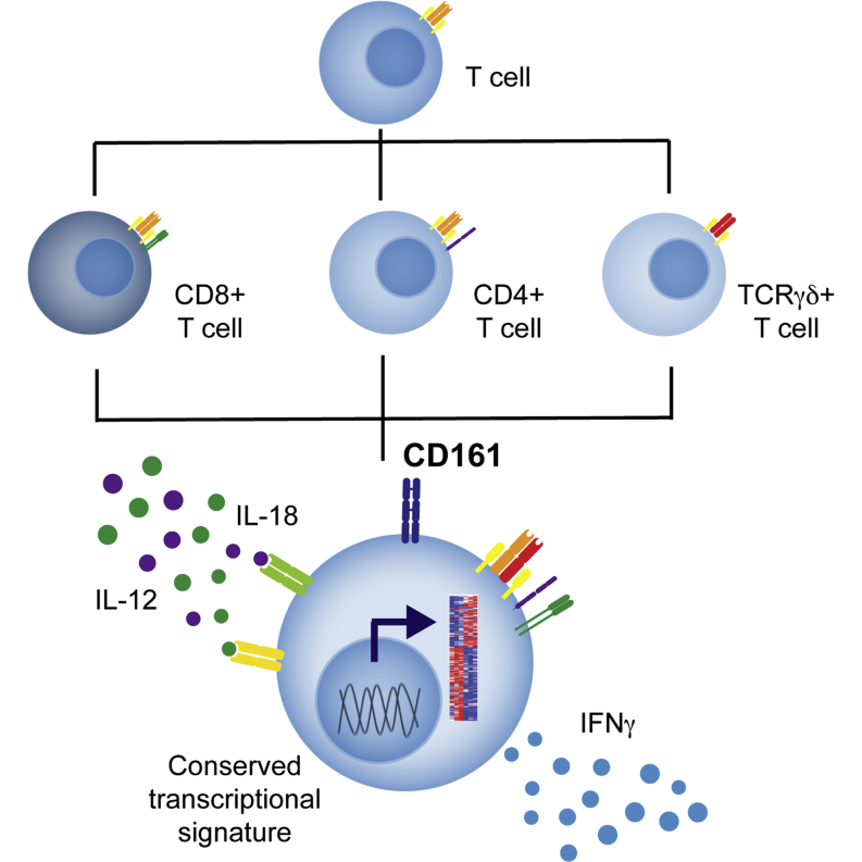
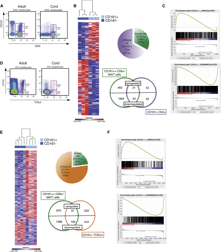
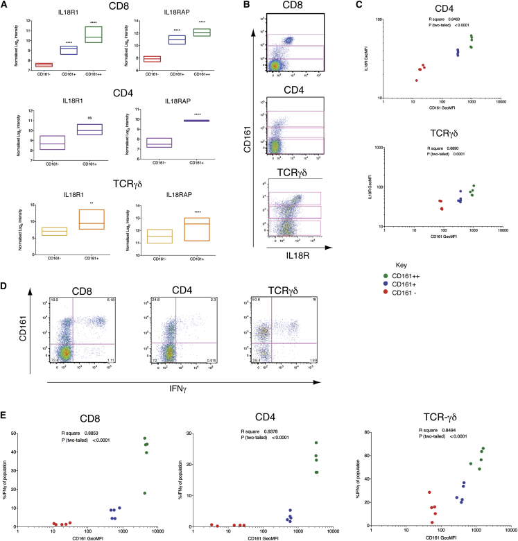
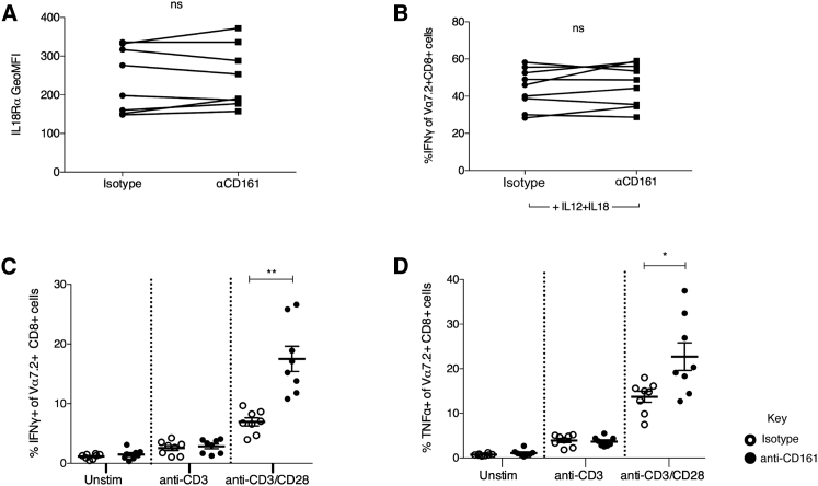
The C-type lectin CD161 is expressed by a large proportion of human T lymphocytes of all lineages, including a population known as mucosal-associated invariant T (MAIT) cells. To understand whether different T cell subsets expressing CD161 have similar properties, we examined these populations in parallel using mass cytometry and mRNA microarray approaches. The analysis identified a conserved CD161++/MAIT cell transcriptional signature enriched in CD161+CD8+ T cells, which can be extended to CD161+ CD4+ and CD161+TCRγδ+ T cells. Furthermore, this led to the identification of a shared innate-like, TCR-independent response to interleukin (IL)-12 plus IL-18 by different CD161-expressing T cell populations. This response was independent of regulation by CD161, which acted as a costimulatory molecule in the context of T cell receptor stimulation. Expression of CD161 hence identifies a transcriptional and functional phenotype, shared across human T lymphocytes and independent of both T cell receptor (TCR) expression and cell lineage.


Figures

None
Graphical abstract
Figure 1
Figure 1
Polyclonal Vα7.2−CD161++ CD8+ T Cells Are Not Activated in an MR1-Dependent Manner but, like MAIT Cells, Respond to Stimulation by IL-12+IL-18 (A) Percentage Vα7.2+ of CD161++CD8+ T cells (histogram) and percentage CD161++ of CD8+ T cells (red point) in cord (n = 3), 24 month (n = 8), and adult blood (n = 8). Representative flow cytometry plots shown above. (B) Comparison of TCR Vβ usage by Vα7.2+ and Vα7.2− CD161++ CD8+ T cells using a TCR Vβ antibody panel. (C) Proportional Vα (38 sequences) and Vβ (129 sequences) usage by Vα7.2− CD161++CD8+ T cells as determined by single-cell TCR sequencing (n = 3). (D) Percentage of IFNγ expression by Vα7.2+ or Vα7.2− CD161++ CD8+ T cell subsets cocultured with THP1 cells exposed to E. coli in the presence or absence of anti-MR1 (10 μg/ml) or corresponding isotype control (n = 11). ∗∗p < 0.01; one-way ANOVA with Dunnett’s multiple comparisons test compared to E. coli alone. (E) Percentage of IFNγ expression by sorted Vα7.2+ or Vα7.2− CD161++CD8+ T cells incubated overnight with IL-12 + IL-18 at 50 ng/ml (n = 9). ns, not significant by paired t test. (F) Transcription factor expression as determined by flow cytometry. Graphs show GeoMFI of RORγt (left) and PLZF (right) by conventional CD8+ T cells (CD3+CD8+ cells excluding CD161++) and Vα7.2+ or Vα7.2− CD161++ CD8+ T cells (n = 5). ∗∗∗∗p < 0.0001; ∗∗p < 0.01; one-way ANOVA with Tukey’s multiple comparison test. Representative histograms show isotype (gray), Vα7.2+ (green line), and Vα7.2− (red) for each transcription factor. All data are represented as mean ± SEM. See also Figure S1.
Figure 2
Figure 2
CyTOF and Principal-Component Analysis Reveals CD161++ Cells to Be Unique among CD8+ T Cells (A) Representative flow cytometry plot of the division of CD8+ T cells into three subsets based upon expression of CD161. (B) Data generated for PMA + ionomycin-stimulated CD8+ T cells were analyzed by principal-component analysis (PCA). The percent variation explained is plotted for each component (bars) and cumulatively (line), and the PCA parameter loadings (weighting coefficients) for the first four components are shown. (C) Stimulated CD8+ T cells from one representative donor are plotted on the first three components as a bulk population (black) and for the three subsets: CD161++ (green); CD161+ (blue); and CD161− (red). (D) Density plots of bulk-stimulated CD8+ T cells are plotted on component 1 and component 4 and are overlaid with contour density of plots of CD161++ (green), CD161+ (blue), and CD161− (red) cells.
Figure 3
Figure 3
CD161+CD8+ T Cells Share a CD161++ Transcriptional Signature and Phenotypic Profile (A) Heatmap showing 3,025 significantly (p < 0.05) differentially expressed transcripts between CD161++ and CD161− CD8+ T cells in four donors. Subsets clustered by one minus Pearson correlation. (B) Heatmap showing 544 significantly differentially expressed genes between CD161+ and CD161− CD8+ T cells in the same four donors. Subsets clustered by one minus Pearson correlation. (C) Pie chart shows proportion of genes differentially expressed by CD161+ that are shared with CD161++ (79%) when compared to CD161− CD8+ T cells. Venn diagram shows breakdown of upregulated and downregulated genes unique to or shared by each subset. (D) Gene set enrichment summary plots for vsn-normalized CD161+ versus CD161− CD8+ T cell ranked genes and CD161++ upregulated (top) and downregulated (bottom) gene sets. Normalized enrichment score (NES) = 3.79, p < 0.001 upregulated genes; NES = −3.12, p < 0.001 downregulated genes. (E) GeoMFI of CD161 for CD161++ (green), CD161+ (blue), and CD161− (red) CD8+ T cell subsets correlated with GeoMFI of CCR6 (r2 = 0.9143; p < 0.0001), CXCR6 (r2 = 0.9258; p < 0.0001), MDR1 (r2 = 0.9322; p < 0.0001), and IL18Rα (r2 = 0.9554; p < 0.0001); n = 5. Representative flow cytometry plots shown for each.
Figure 4
Figure 4
CD161+CD4+ and CD161+TCRγδ+ T Cells Are Enriched for the CD161++CD8+/MAIT Transcriptional Signature (A) Representative flow cytometry plot showing CD161+CD4+ T cells within adult and cord blood. (B) Heatmap showing 169 significantly differentially expressed genes between CD161+ and CD161− CD4+ T cells in three donors. Subsets clustered by one minus Pearson correlation. Pie chart shows proportion of genes differentially expressed by CD161+CD4+ that are shared with CD161++CD8+ (33%), when compared to their CD161− counterparts. Venn diagram shows breakdown of upregulated and downregulated genes unique to or shared by each subset. (C) Gene set enrichment summary plots for vsn-normalized CD161+ versus CD161− CD4+-T-cell-ranked genes and CD161++CD8+ upregulated (left) and downregulated (right) genes. NES = 2.45, p < 0.001 upregulated genes; NES = −3.04, p < 0.001 downregulated genes. (D) Representative flow cytometry plot showing CD161+TCRγδ+ T cells within adult and cord blood. (E) Heatmap showing 828 significantly differentially expressed genes between CD161+ and CD161− TCRγδ+ T cells in four donors. Subsets clustered by one minus Pearson correlation. Pie chart shows proportion of genes differentially expressed by CD161+TCRγδ+ that are shared with CD161++CD8+ (21%), when compared to their CD161− counterparts. Venn diagram shows breakdown of upregulated and downregulated genes unique to or shared by each subset. (F) Gene set enrichment summary plots for vsn-normalized CD161+ versus CD161− TCRγδ+-T-cell-ranked genes and CD161++CD8+ upregulated (left) and downregulated (right) genes. NES = 2.78, p < 0.001 upregulated genes; NES = −2.62, p < 0.001 downregulated genes.
Figure 5
Figure 5
CD161-Positive T Cell Subsets Express Elevated Levels of IL18Rα and Respond to Stimulation by IL-12+IL-18 (A) Relative expression levels of IL18R subunits by CD161-positive and CD161-negative sorted T cells and statistical significance from mRNA expression analyses after normalization and correction for multiple testing. ∗∗p < 0.01; ∗∗∗∗p < 0.0001. Floating bars show minimum and maximum values, with a line at the mean. (B) Representative flow cytometry plots for IL18Rα expression in each T cell lineage, revealing CD161++, CD161+, and CD161− subsets in each (gated). (C) GeoMFI of CD161 for CD161++ (green), CD161+ (blue), and CD161− (red) subsets in CD4+ and TCRγδ+ T cells correlated with GeoMFI of IL18Rα. CD4 r2 = 0.8463, p < 0.0001; TCRγδ r2 = 0.6890, p < 0.0001 (n = 5). (D) PBMCs were stimulated overnight with 50 ng/ml IL-12+IL-18. Representative flow cytometry plots for IFNγ expression by gated CD8+, CD4+, and TCRγδ+ CD3+ live lymphocytes. (E) GeoMFI of CD161 for CD161++ (green), CD161+ (blue), and CD161− (red) subsets in CD8+, CD4+, and TCRγδ+ T cells correlated with percentage of each subset expressing IFNγ in response to overnight stimulation with IL-12+IL-18. CD8 r2 = 0.8853, p < 0.0001; CD4 r2 = 0.9378, p < 0.0001; TCRγδ r2 = 0.8494, p < 0.0001 (n = 5). See also Figure S2.
Figure 6
Figure 6
CD161 Regulates TCR-Dependent, but Not TCR-Independent, Responses (A) GeoMFI of IL18Rα on Vα7.2+CD8+CD3+ cells incubated for 18 hr with biotin beads coated with either IgG2a isotype or anti-CD161. ns, not significant by paired t test (n = 9). (B) Percentage of Vα7.2+CD8+CD3+ cells expressing IFNγ when cultured with 50 ng/ml IL-12+IL-18 for 18 hr in the presence of biotin beads coated with either IgG2a isotype or anti-CD161. ns, not significant by paired t test (n = 9). (C and D) Percentage of Vα7.2+CD8+CD3+ cells expressing IFNγ (C) or TNF-α (D) when cultured overnight with TCR-stimulating biotin beads, coated either with anti-CD3 or anti-CD3 + anti-CD28, in combination with IgG2a isotype (open circles) or anti-CD161 (filled circles). p < 0.05 and ∗∗p < 0.01 by repeated-measures one-way ANOVA (n = 8). Data are represented as mean ± SEM. See also Figure S3.

References

    1. Aldemir H., Prod’homme V., Dumaurier M.-J., Retiere C., Poupon G., Cazareth J., Bihl F., Braud V.M. Cutting edge: lectin-like transcript 1 is a ligand for the CD161 receptor. J. Immunol. 2005;175:7791–7795. - PubMed
    1. Annibali V., Ristori G., Angelini D.F., Serafini B., Mechelli R., Cannoni S., Romano S., Paolillo A., Abderrahim H., Diamantini A. CD161(high)CD8+T cells bear pathogenetic potential in multiple sclerosis. Brain. 2011;134:542–554. - PubMed
    1. Annunziato F., Cosmi L., Santarlasci V., Maggi L., Liotta F., Mazzinghi B., Parente E., Filì L., Ferri S., Frosali F. Phenotypic and functional features of human Th17 cells. J. Exp. Med. 2007;204:1849–1861. - PMC - PubMed
    1. Bai A., Moss A., Kokkotou E., Usheva A., Sun X., Cheifetz A., Zheng Y., Longhi M.S., Gao W., Wu Y., Robson S.C. CD39 and CD161 Modulate Th17 Responses in Crohn’s Disease. J. Immunol. 2014;193:3366–3377. - PMC - PubMed
    1. Battistini L., Borsellino G., Sawicki G., Poccia F., Salvetti M., Ristori G., Brosnan C.F. Phenotypic and cytokine analysis of human peripheral blood gamma delta T cells expressing NK cell receptors. J. Immunol. 1997;159:3723–3730. - PubMed

Publication types

MeSH terms

Substances

Associated data