

Ameloblastic fibro-odontoma
- PMID: 25437658
- PMCID: PMC4276268
- DOI: 10.1016/j.ijscr.2014.11.025
Ameloblastic fibro-odontoma
Abstract
Introduction: Ameloblastic fibro-odontoma (AFO) is a quite rare, mixed odontogenic tumour generally seen in the early stages of life. Frequent signs of this tumour are asymptomatic swelling, delayed tooth eruption and mixed radiological appearance within well-defined borders. Management of the lesion includes enucleation of the tumour and long-term follow-up.
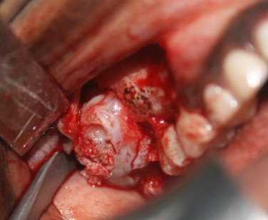
Presentation of case: A 10-year-old girl was referred to our oral and maxillofacial surgery clinic with an incidental radiological finding of radiopaque mass in the posterior region of maxilla. OPG showed unerupted tooth bud of upper right second molar and was being prevented from eruption by the odontome. Under general anaesthesia, the lesion was enucleated and the permanent right upper second molar tooth bud removed.
Discussion: Mixed odontogenic tumours are a group of rare and interesting lesions which can mislead the clinician to variety of differential diagnosis. Adequate clinical and radiological investigations, proper surgical excison, accurate histopathological diagnosis, and long term follow up will ensure the right treatment plan for the patient.
Conclusion: The possibility of a mixed rare tumour should be kept in mind by the clinician where they deal with the swellings of posterior maxilla in children. Histological assessment revealed a final diagnosis of ameloblastic fibro-odontoma.
Keywords: Ameloblastic fibro-odontoma; Odontome; Posterior maxillary swelling mixed odontogenic tumours.
Copyright © 2014 The Authors. Published by Elsevier Ltd.. All rights reserved.
Figures


References
-
- Barnes L., Eveson J.W., Reichart P., Sidransky D. World Health Organization Classification of Tumours, IARC Press; Lyon: 2005. Pathology and genetics. Head and neck tumors.
-
- Furst I., Pharoah M., Phillips J. Recurrence of an ameloblastic fibro-odontoma in a 9-year-old boy. J Oral Maxillofac Surg. 1999;57:620–623. - PubMed
-
- Buchner A., Merrell P.W., Carpenter W.M. Relative frequency of central odontogenic tumors: a study of 1,088 cases from Northern California and comparison to studies from other parts of the world. J Oral Maxillofac Surg. 2006;64:1343–1352. - PubMed
-
- Takeda Y. Ameloblastic fibroma and related lesions: current pathologic concept. Oral Oncol. 1999;35:535–540. - PubMed
-
- Chang H., Precious D.S., Shimizu M.S. Ameloblastic fibro-odontoma: a case report. J Can Dent Assoc. 2002;68:243–246. - PubMed
LinkOut - more resources
Full Text Sources
Other Literature Sources
Research Materials
