Review
. 2014 Apr 10;3(2):280-308.
doi: 10.3390/pathogens3020280.

Leptospiral pathogenomics

Affiliations
Review

Leptospiral pathogenomics

Jason S Lehmann et al. Pathogens. .

Abstract

Leptospirosis, caused by pathogenic spirochetes belonging to the genus Leptospira, is a zoonosis with important impacts on human and animal health worldwide. Research on the mechanisms of Leptospira pathogenesis has been hindered due to slow growth of infectious strains, poor transformability, and a paucity of genetic tools. As a result of second generation sequencing technologies, there has been an acceleration of leptospiral genome sequencing efforts in the past decade, which has enabled a concomitant increase in functional genomics analyses of Leptospira pathogenesis. A pathogenomics approach, by coupling of pan-genomic analysis of multiple isolates with sequencing of experimentally attenuated highly pathogenic Leptospira, has resulted in the functional inference of virulence factors. The global Leptospira Genome Project supported by the U.S. National Institute of Allergy and Infectious Diseases to which key scientific contributions have been made from the international leptospirosis research community has provided a new roadmap for comprehensive studies of Leptospira and leptospirosis well into the future. This review describes functional genomics approaches to apply the data generated by the Leptospira Genome Project towards deepening our knowledge of virulence factors of Leptospira using the emerging discipline of pathogenomics.


Figures

Figure 1
Figure 1
Taxonomy of the Genus Leptospira. A phylogenetic tree of full-length 16S rRNA sequences showing the relatedness of the 21 recognized leptospiral species. The putative Clade C has been detected in Peruvian surface waters by qPCR [23] and includes strains/species of unknown pathogenicity. For brevity, the Leptospira genus name has been omitted. Key inferred evolutionary events as indicated by whole genome comparisons are shown. Prophages have been detected in L. interrogans, L. licerasiae, and L. biflexa, but not L. borgpetersenii. Of these, only in L. interrogans have the putatively antiviral “clustered regularly interspaced short palindromic repeats” (CRISPR) elements been identified. By contrast, L. licerasiae and L. biflexa have an expanded repertoire of type II and type III toxin-antitoxin systems, which could have anti-phage activity. Of the pathogenic species, only the more virulent Group I pathogens have genes for putative virulence proteins belonging to the paralogous family matching Pfam model PF07598, which we suggest help determine tissue-specific colonization. The outgroup is the closely related spirochete Leptonema illini. For the published genomes, complete genomes are available for six strains of three species (*) and high-quality draft genomes are currently available in GenBank for three strains (**). Genomes representing the remaining Leptospira species are available in GenBank (manuscript in preparation).
Figure 2
Figure 2
Blast Ring Image Generator (BRIG) [115] plot showing whole genome comparison of L. interrogans, L. borgpetersenii, L. licerasiae and L. biflexa. Track 1 (innermost; reference genome): L. interrogans Lai 56601; Track 2: GC%; Track 3: GC Skew; Track 4: L. interrogans Lai IPAV; Track 5: L. interrogans Copenhageni L1-130; Track 6: L. borgpetersenii Hardjo (JB197 and L550); Group II (Track 7): L. licerasiae Varillal (VAR010 and MMD0835); and Track 8: L. biflexa Patoc (Ames and Paris) showing conserved proteins with amino acid similarity of ≥50%. The locations of two common IS elements in Lai 56601: ISlin1 (red) and IS1533 (blue) are shown. The solid black line shows the inverted region of the Lai 56601 genome (with respect to Copenhageni L1-130); the two IS elements believed to have mediated the inversion have been designated with an asterisk (*). The location of 54-kb GI in Lai 56601, capable of excising from the chromosome and replicating as a plasmid [101], is shown. Homologous coding regions have been color-coded based on %identity as indicated by the key. All Leptospira contain both the CI and CII replicons; whereas only L. biflexa contains a large plasmid designated p74 (not shown).
Figure 3
Figure 3
Genetic organization and alternate location of L. licerasiae rfb locus. (A) Comparison of the rfb locus of L. licerasiae strain VAR010 and homologous region in L. interrogans serovar Copenhageni; (B) Comparison of the rfb locus of L. interrogans Copenhageni and homologous region in L. licerasiae strain VAR10. Yellow shaded boxes mark the locations of the O-antigen regions. CDSs are labeled by locus identifier and colored by functional role categories as noted in the boxed key. Gene symbols, when present, are noted above their respective genes in bold italics. Reproduced from [87].
Figure 4
Figure 4
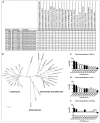
Distribution and expression profile of PF07598 gene family (encoding putatively named virulence modulating or VM proteins). (A) Distribution of PF07598 across the genus Leptospira; P, pathogen; I, intermediate; S, saprophyte. (B) Unrooted phylogenetic tree of the VM protein family including sequences from Helicobacter spp. and Bartonella bacilliformis. Node labels represent support from 500 bootstrap replicates. (CE) Transcript levels of each PF07598 paralog were assessed by real time, reverse transcriptase quantitative PCR of blood, liver and kidney extracts four days after hamster infection and compared to log phase in vitro cultured Leptospira; expressed as the log2 of the fold change between the two conditions. Solid bars indicate proteins potentially extracellular proteins. Data represented are the mean ± SEM of three independent experiments (n = 7 animals). Reproduced from reference [69].
Figure 5
Figure 5
Venn diagram showing the distribution of shared and unique proteins separated by leptospiral species. Two strains from each species were used for these comparisons. Only proteins present in both strains of a given species are shown. Reproduced from [87].

References

    1. Bharti A.R., Nally J.E., Ricaldi J.N., Matthias M.A., Diaz M.M., Lovett M.A., Levett P.N., Gilman R.H., Willig M.R., Gotuzzo E. Leptospirosis: a zoonotic disease of global importance. Lancet Infect. Dis. 2003;3:757–771. doi: 10.1016/S1473-3099(03)00830-2. - DOI - PubMed
    1. Abela-Ridder B., Sikkema R., Hartskeerl R.A. Estimating the burden of human leptospirosis. Int. J. Antimicrob. Agents. 2010;36(Suppl. 1):S5–S7. doi: 10.1016/j.ijantimicag.2010.06.012. - DOI - PubMed
    1. Ko A.I., Galvao Reis M., Ribeiro Dourado C.M., Johnson W.D., Jr., Riley L.W. Urban epidemic of severe leptospirosis in Brazil. Salvador Leptospirosis Study Group. Lancet. 1999;354:820–825. doi: 10.1016/S0140-6736(99)80012-9. - DOI - PubMed
    1. Gouveia E.L., Metcalfe J., de Carvalho A.L., Aires T.S., Villasboas-Bisneto J.C., Queirroz A., Santos A.C., Salgado K., Reis M.G., Ko A.I. Leptospirosis-associated severe pulmonary hemorrhagic syndrome, Salvador, Brazil. Emerg. Infect. Dis. 2008;14:505–508. doi: 10.3201/eid1403.071064. - DOI - PMC - PubMed
    1. Zaki S.R., Shieh W.J., The Epidemic Working Group at Ministry of Health in Nicaragua Leptospirosis associated with outbreak of acute febrile illness and pulmonary haemorrhage, Nicaragua, 1995. Lancet. 1996;347:535–536. doi: 10.1016/S0140-6736(96)91167-8. - DOI - PubMed

LinkOut - more resources